| Cardiology Research, ISSN 1923-2829 print, 1923-2837 online, Open Access |
| Article copyright, the authors; Journal compilation copyright, Cardiol Res and Elmer Press Inc |
| Journal website https://cr.elmerpub.com |
Original Article
Volume 17, Number 6, December 2026, pages 664-669

Virtual Assessment of Coronary Microcirculation Before and After Rotational Atherectomy
Gueladio Konea, Brahim Baalaa, Alexandre Canvillea, Matthieu Godina, Quentin Landolffa, b
aDepartment of Interventional Cardiology, Clinique Saint-Hilaire, Rouen, France
bCorresponding Author: Quentin Landolff, Department of Interventional Cardiology, Clinique Saint-Hilaire, 76000 Rouen, France
Manuscript submitted June 18, 2026, accepted September 24, 2026, published online October 24, 2026
Short title: Coronary Microcirculation After RA
doi: https://doi.org/10.14740/cr2276
| Abstract | ▴Top |
Background: Calcified coronary lesions remain a major challenge during percutaneous coronary intervention (PCI). Rotational atherectomy (RA) (Boston Scientific) is a well-established tool used to modify calcified plaques and facilitate stent delivery. Despite its mechanical benefits, RA may adversely affect coronary microcirculation through distal plaque debris embolization. Angiography-derived microvascular resistance (AMR) (Pulse Medical) is a wire-free index correlated with invasive indices such as the index of microcirculatory resistance (IMR), which allows assessment of microcirculatory resistance directly from angiographic images, without a pressure wire or adenosine. The objective of this study is to evaluate changes in AMR before and after RA and to identify clinical, angiographic, and procedural predictors of postprocedural coronary microvascular dysfunction (CMD).
Methods: We performed a retrospective single-center observational study including all patients who underwent RA between January 2023 and December 2024 at Clinique Saint-Hilaire in Rouen, France. AMR was measured before and after RA using the AngioPlus software (Pulse Medical). CMD was defined as postprocedural AMR > 250 mm Hg·s/m.
Results: Of 49 RA procedures, 40 patients were included. AMR increased in all cases but CMD occurred in 42.5% of patients (n=17). Diabetes mellitus and reduced left ventricular ejection fraction (LVEF) were significantly more prevalent in the CMD group. Long coronary artery lesions (> 20 mm), larger plaque burden, and prolonged burr run time were associated with higher AMR values. Mean AMR rose from 176 to 315 mm Hg·s/m in the CMD group, versus 177 to 201 mm Hg·s/m in the non-CMD group.
Conclusions: Performing RA affects the coronary microcirculation, especially in cases of diabetes mellitus and reduced LVEF.
Keywords: Rotablator; Angiography-derived microvascular resistance; Coronary microvascular dysfunction
| Introduction | ▴Top |
Coronary artery disease (CAD) is a major cause of morbidity and mortality worldwide. Calcified coronary lesions represent around 30% of coronary lesions, particularly prevalent in elderly and diabetic populations. Percutaneous coronary interventions (PCI) on these lesions can be challenging, impairing balloon dilatation, limiting stent expansion, and increasing the risks of stent restenosis and thrombosis.
In this context, rotational atherectomy (RA) (Boston Scientific) represents a well-established lesion preparation tool, modifying heavily calcified plaques, facilitating stent delivery and expansion. However, this technique may release plaque debris that can obstruct the distal microcirculation, causing slow-flow or no-reflow phenomena and potentially reducing left ventricular function.
The assessment of coronary microcirculation traditionally relied on invasive wire-based indices such as coronary flow reserve (CFR) or the index of microcirculatory resistance (IMR) measured by Coroflow software (Abbott), which require a pressure wire and pharmacologically induced hyperemia. An IMR value above 25 is accepted for microvascular dysfunction. Although validated, these methods still are underused due to their logistic complexity and cost. Recently, angiography-derived microvascular resistance (AMR) measured by AngioPlus software (Pulse Medical) emerged as a wire-free, rapid, and reproducible alternative that correlates well with IMR (Fig. 1), with a postprocedural AMR ≥ 250 mm Hg·s/m being associated with microvascular dysfunction.
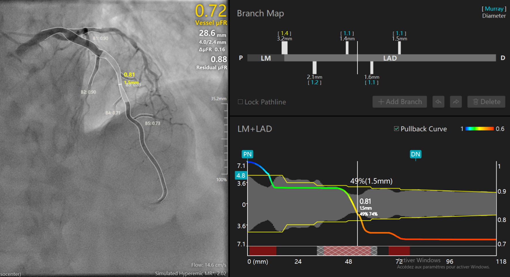 Click for large image | Figure 1. Example of microFR and AMR measurements with AngioPlus (Pulse Medical). Left panel: angiogram with the reconstructed LM–LAD. B1 to B5 designate the side branches identified along the vessel, with the microFR value computed at the level of each branch. The yellow value (0.81; 1.5 mm) indicates the microFR and the minimal lumen diameter at the site of the lesion. Upper-right values: vessel microFR (0.72), lesion length (28.6 mm), proximal/distal reference diameters (4.0/2.4 mm), pressure drop across the lesion = 0.16 (it is the variation of microFR after treatment ΔmicroFR) and predicted residual microFR after treatment of the lesion (0.88). Lower values: estimated contrast flow velocity (14.6 cm/s) and simulated hyperemic microvascular resistance, AMR (2.02 mm Hg·s/cm, corresponding to 202 mm Hg·s/m). Right upper panel (branch map): schematic representation of the LM and LAD from proximal (P) to distal (D), showing the position and diameter of each side branch. Right lower panel (LM + LAD pullback curve): virtual microFR pullback along the vessel. The vertical line marks the minimal lumen diameter site (1.5 mm; 49% diameter stenosis and 74% area stenosis; microFR, 0.81). AMR: angiography-derived microvascular resistance; LAD: left anterior descending artery; LM: left main coronary artery; microFR: Murray-law-based quantitative flow ratio. |
The aim of this study was to evaluate the impact of RA on coronary microcirculation using AMR and to identify clinical, angiographic, and procedural factors associated with postprocedural microvascular dysfunction. We hypothesized that RA induces a measurable increase in AMR, reflecting coronary microvascular injury.
| Materials and Methods | ▴Top |
Study design and population selection
This retrospective, single-center, non-randomized study was conducted at the Department of Interventional Cardiology, Clinique Saint-Hilaire, Rouen, France from January 2023 to December 2024. In this period, 49 RA procedures were performed. Inclusion criteria were age > 18 years; at least one severely calcified coronary lesion treated with RA; angiographic image quality sufficient for AMR analysis (acquisition rate ≥ 7.5 frames/s) both before and after the procedure, and availability of 1-year clinical follow-up through the France PCI registry. Exclusion criteria included prior bypass grafting of the target vessel, poor angiographic image quality, and ST-elevation myocardial infarction (STEMI) (Fig. 2).
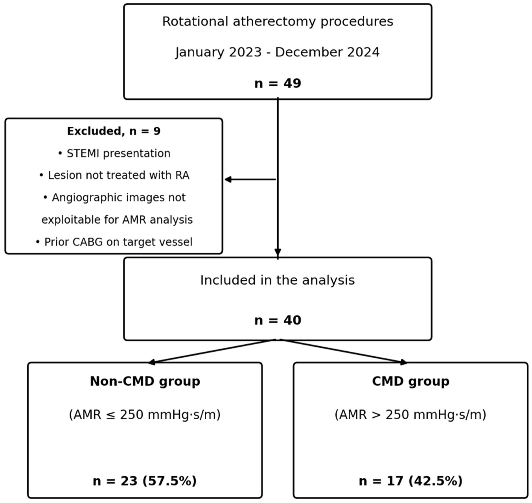 Click for large image | Figure 2. Flow chart of the study. AMR: angiography-derived microvascular resistance; CABG: coronary artery bypass grafting; CMD: coronary microvascular dysfunction; RA: rotational atherectomy; STEMI: ST-elevation myocardial infarction. |
We applied these exclusion criteria because of factors that could compromise the validity of AMR analysis, such as distal coronary flow disturbance in patients with coronary artery bypass grafting (CABG), suboptimal angiographic image quality (non-Thrombolysis in Myocardial Infarction (TIMI) 3 flow, tortuosity), and transient microvascular injury in patients with STEMI.
Procedural protocol
RA was performed with the Rotablator system (Boston Scientific, Marlborough, MA, USA) by an experienced operator, according to standardized departmental practice and current recommendations. After lesion crossing with a dedicated RotaWire, a burr size of 1.25–1.75 mm was selected, targeting a burr-to-artery ratio close to 0.5. Burr rotation was initiated at low speed (60,000–70,000 revolutions per minute (rpm)) before engaging the lesion, then increased to a working speed of 130,000–180,000 rpm (up to ≥ 180,000 rpm when dilatation remained incomplete); each run was limited to less than 30 s. Anticoagulation followed guidelines [1].
AMR analysis
The analysis was performed with AngioPlus software (Pulse Medical) using a validated algorithm that estimates distal pressure and hyperemic velocity from angiographic flow. AMR was measured before and after RA. Values ≥ 250 mm Hg·s/m after RA were considered abnormal. A threshold based on a validated cutoff was compared with invasively measured coronary microcirculatory resistance indices [2, 3].
AMR was calculated as follows: AMR = Pd/Vhyp = Pa × microFR/Vhyp [4], where Pd is the distal coronary pressure (mm Hg), Pa is the mean aortic pressure (mm Hg) recorded at the tip of the guiding catheter; microFR is the Murray-law-based quantitative flow ratio, and Vhyp is the hyperemic mean flow velocity (m/s) derived from the contrast. AMR is therefore expressed in mm Hg·s/m.
Because AMR relies on accurate three-dimensional vessel reconstruction, its reliability can be affected by suboptimal postprocedural image quality (e.g., contrast staining, residual dissection flap, or transiently reduced flow). All angiograms included in the analysis, before and after RA, met the same quality criteria; studies not meeting this standard were excluded. We acknowledge that a dedicated technical validation of AngioPlus specifically after RA has not been performed to date and remains needed.
Data collection
Clinical, angiographic, and procedural data were collected retrospectively. Statistical analyses were performed using Microsoft Excel and ODK Central. ODK Central is a platform used as a standardized and secure electronic data-capture server.
Statistical analysis
Continuous variables are expressed as mean ± standard deviation and were compared between the coronary microvascular dysfunction (CMD) and non-CMD groups using Student’s t-test. The paired difference between preprocedural and postprocedural AMR (ΔAMR) was assessed using a paired t-test
Follow-up
One-year clinical follow-up data were collected from the France PCI registry.
Ethics
The Institutional Review Board (IRB) approved the study. This study was conducted in compliance with the ethical standards of the responsible institution on human subjects as well as with the Helsinki Declaration.
| Results | ▴Top |
Population characteristics
Mean age was 75 years, and 87% were male. Among the cardiac risk factors, diabetes mellitus was present in 47% overall and was more prevalent in in the CMD group (70% vs 30%).
Reduced left ventricular ejection fraction (LVEF) was more common in the CMD group (41% vs 17%). Other cardiovascular risk factors, comorbidities, and clinical presentation were comparable between groups (Table 1).
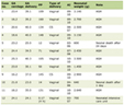 Click to view | Table 1. Clinical Characteristics |
Procedural findings
Radial access was used in 97% of cases.
Target coronary artery lesions were located in the left anterior descending artery (LAD) in 65% of cases, the right coronary artery (RCA) in 27%, and the left circumflex artery (LCX) in 8%. Left main coronary artery lesions were present in 10% of the overall population, without significant difference between the two groups.
Lesions longer than 20 mm were more prevalent in the CMD group (70%). Multivessel disease was frequent in both groups.
A 1.5-mm burr was used in the majority of cases and comparably in both groups. The 1.75-mm burr was used exclusively in the CMD group (two patients) (Table 2).
Click to view | Table 2. Procedural Characteristics |
Microcirculation result findings
AMR increased in all patients after RA (100%), rising from 177 to 201 mm Hg·s/m in the non-CMD group and from 176 to 315 mm Hg·s/m in the CMD group. This result suggests that AMR may serve as a marker of coronary microvascular injury (Table 1).
Complications
Coronary artery dissections occurred only in the CMD group (17%).
Follow-up data were available for all 40 patients through the France PCI registry. During the 1-year follow-up, three long-term events were recorded from the France PCI registry, all in the non-dysfunction group: one death, one myocardial infarction, and one stroke.
| Discussion | ▴Top |
This study demonstrates that RA is consistently associated with a rise in coronary microvascular resistance with significant variation according to patient and lesion characteristics and with CMD occurring in 42.5% of patients.
Patients who developed CMD after RA more frequently presented with cardiovascular risk factors such as diabetes mellitus and current smoking. This is consistent with the results of Williams et al, who demonstrated that chronic hyperglycemia and high glycated hemoglobin levels are known to impair endothelial function and red cell rheology, both of which could plausibly contribute to increased resistance in the coronary microcirculation [5]. In our study, diabetes mellitus was found to be an important predictive factor for CMD. Other classical risk factors, such as hypertension and dyslipidemia, were numerically more frequent in the CMD group but did not reach statistical significance.
Procedural characteristics, particularly the length of the coronary artery lesion treated with RA, were identified as major determinants of CMD in our study. It often reflects the presence of more severe calcification and a larger plaque burden, which are responsible for an increased production of debris during RA.
Wang et al reported CMD in 45.8% of cases after RA, particularly among patients with long or multivessel coronary artery lesions [6]. In our study, 70% of patients with CMD after RA had coronary artery lesions longer than 20 mm. This comparison should be interpreted cautiously, as the two cohorts differ in sample size and in the microcirculatory index used: Wang et al [6] assessed an angiography-derived IMR rather than AMR.
Several techniques are available to assess coronary microvascular resistance. Invasive approaches such as IMR remain the reference due to their diagnostic accuracy, particularly when the IMR value exceeds 25 [7]. However, IMR presents certain limitations, including the use of hyperemic agents and pressure wires, which lengthen the procedure and increase its cost. Likewise, cardiac magnetic resonance imaging (MRI) is considered the gold standard for evaluating myocardial perfusion and tissue viability. However, it faces logistical constraints, including limited availability, long examination times, and high costs.
To overcome these limitations, AMR has emerged as a promising alternative. AMR is a virtual software-based tool that requires neither a pressure wire nor adenosine, and it is derived from angiography, allowing a simplified assessment of coronary microvascular resistance. This method offers a more accessible and cost-effective solution [8]. Prior validation work, including studies in patients with chronic coronary syndromes [3] and studies using computational fluid-dynamics models comparable to those underlying AMR [4, 9], has reported good correlations between angiography-derived and invasively measured microcirculatory indices. Nonetheless, dedicated validation of AMR specifically in the post-RA setting is still lacking and represents a priority for future research.
In our study, post–RA AMR was significantly higher in patients with CMD, with mean AMR increasing from 176 to 315 mm Hg·s/m in the CMD group.
Clinical implications
Low-flow and no-reflow during RA are recognized complications secondary to microvascular injury. Strategies to minimize coronary microvascular injury may include minimizing rotational time, using smaller burr sizes, and giving adjunctive vasodilators.
The study is limited by its retrospective design, modest sample size, and lack of direct IMR comparison.
AMR accuracy also depends on angiographic quality.
Limitations
This study has several limitations. It is retrospective, single-center, and non-randomized; the sample size is modest. No direct invasive IMR comparison was available. Although AMR has shown good correlation with IMR in prior validation studies, dedicated technical validation of AngioPlus specifically in the post-RA setting has not been performed. No biological markers of myocardial injury (e.g., troponin) were collected. Given the very small number of events, no formal statistical outcome analysis was performed; we chose to retain these events as a descriptive, hypothesis-generating observation. No comparator group (balloon angioplasty or another calcified-plaque-modification technique) was included. Finally, AMR calculation quality depends on angiographic acquisition quality, which may itself be affected after RA.
Conclusions
RA systematically increases AMR, reflecting coronary artery microvascular stress, with coronary artery dysfunction in nearly half of patients. Diabetes mellitus, reduced LVEF, coronary artery lesion length and prolonged RA duration are key predictors of CMD. AMR is a reliable, wire-free tool for evaluating coronary microcirculation and may enhance decision-making during complex PCI.
Future multicenter prospective trials should compare AMR with IMR and assess long-term prognostic value. Integration of AMR in PCI workflows will allow better patient selection and improve outcomes in complex calcified CAD.
Acknowledgments
None to declare.
Financial Disclosure
None to declare.
Conflict of Interest
None to declare.
Informed Consent
Given the retrospective design of the study and in accordance with French regulations governing research using existing data, each participant received an information letter describing the study and was given the opportunity to object to the use of their data. No participant returned an objection or declined participation.
Author Contributions
GK, BB, AC, MG, QL performed the procedures. GK wrote the manuscript. QL reviewed the manuscript.
Data Availability
The data supporting the findings of this study are available from the corresponding author upon reasonable request.
Abbreviations
AMR: angiography-derived microvascular resistance; CAD: coronary artery disease; CFR: coronary flow reserve; IMR: index of microcirculatory resistance; LAD: left anterior descending artery; LCX: left circumflex artery; LVEF: left ventricular ejection fraction; MRI: magnetic resonance imaging; microFR: Murray-law-based quantitative flow ratio; PCI: percutaneous coronary interventions; RA: rotational atherectomy; RCA: right coronary artery
| References | ▴Top |
- Barbato E, Carrie D, Dardas P, Fajadet J, Gaul G, Haude M, Khashaba A, et al. European expert consensus on rotational atherectomy. EuroIntervention. 2015;11(1):30-36.
doi pubmed - Tu S, Ding D, Chang Y, Li C, Wijns W, Xu B. Diagnostic accuracy of quantitative flow ratio for assessment of coronary stenosis significance from a single angiographic view: a novel method based on bifurcation fractal law. Catheter Cardiovasc Interv. 2021;97uppl 2):1040-1047.
doi pubmed - Qiu Z, Wang Y, Liu Y, Zhou Z, Wang Z. Diagnostic value of angiography-derived index of microcirculatory resistance (AMR) for coronary microcirculatory dysfunction (CMD) and its prognostic significance in patients with chronic coronary syndromes in the smoking population. Medicine (Baltimore). 2024;103(6):e37022.
doi pubmed - Fan Y, Fezzi S, Sun P, Ding N, Li X, Hu X, Wang S, et al. In vivo validation of a novel computational approach to assess microcirculatory resistance based on a single angiographic view. J Pers Med. 2022;12(11).
doi pubmed - Williams A, Bissinger R, Shamaa H, Patel S, Bourne L, Artunc F, Qadri SM. Pathophysiology of red blood cell dysfunction in diabetes and its complications. Pathophysiology. 2023;30(3):327-345.
doi pubmed - Wang B, Gao Y, Zhao Y, Jia P, Han J, Li H, Zhang Y, et al. Prognostic value of angiography-derived index of microcirculatory resistance in patients with coronary artery disease undergoing rotational atherectomy. Rev Cardiovasc Med. 2023;24(5):131.
doi pubmed - Fearon WF, Kobayashi Y. Cut-off values for invasive microvascular dysfunction assessments. Circulation. 2017;136(6):520-533.
- Mejia-Renteria H, Lee JM, Choi KH, Lee SH, Wang L, Kakuta T, Koo BK, et al. Coronary microcirculation assessment using functional angiography: development of a wire-free method applicable to conventional coronary angiograms. Catheter Cardiovasc Interv. 2021;98(6):1027-1037.
doi pubmed - Li J, Gong Y, Wang W, Yang Q, Liu B, Lu Y, Xu Y, et al. Accuracy of computational pressure-fluid dynamics applied to coronary angiography to derive fractional flow reserve: FLASH FFR. Cardiovasc Res. 2020;116(7):1349-1356.
doi pubmed
This article is distributed under the terms of the Creative Commons Attribution 4.0 International License (CC BY 4.0), which permits unrestricted use, distribution, and reproduction in any medium, including commercial use, provided the original work is properly cited.
Cardiology Research is published by Elmer Press Inc.