Cardiology Research, ISSN 1923-2829 print, 1923-2837 online, Open Access
Article copyright, the authors; Journal compilation copyright, Cardiol Res and Elmer Press Inc
Journal website https://cr.elmerpub.com

Case Report

Volume 15, Number 6, December 2024, pages 472-476


The Mechanism and Management of Pneumopericardium Caused by Right Ventricular Lead Perforation

Figures

Figure 1.
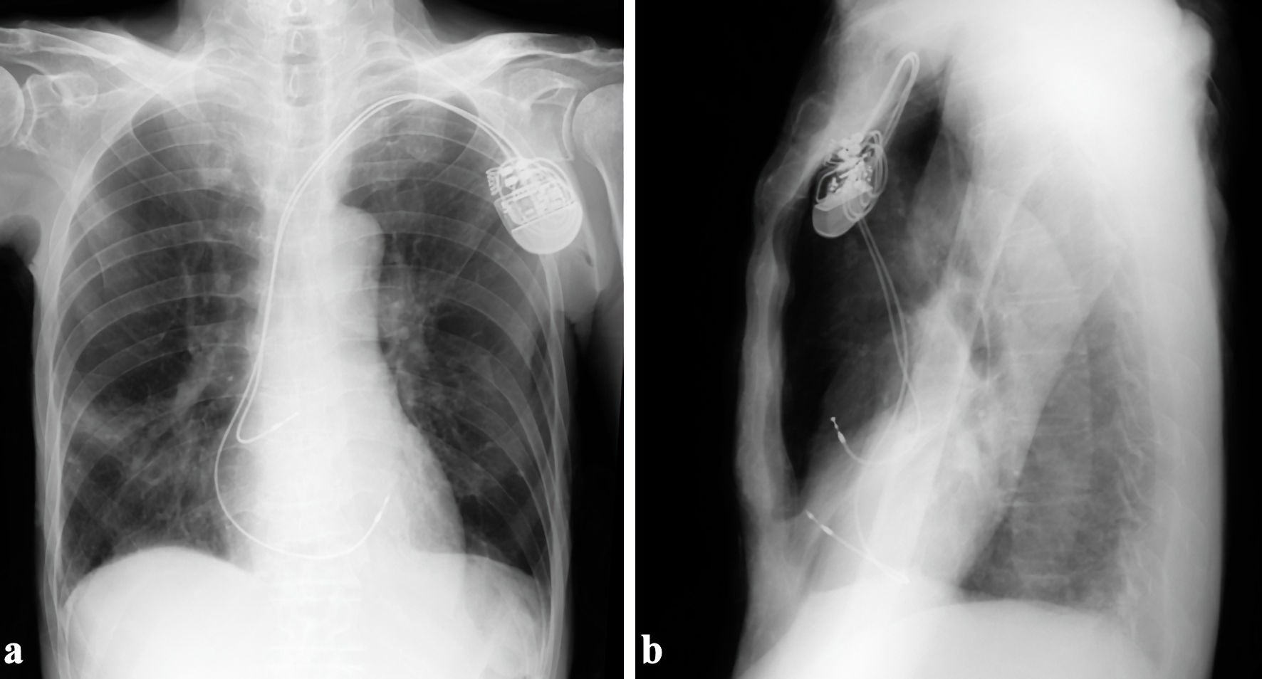
Figure 1. The ventricular active-fixation lead was placed in the anterior right ventricular free wall. (a) Anteroposterior view. (b) Right-left view.
Figure 2.
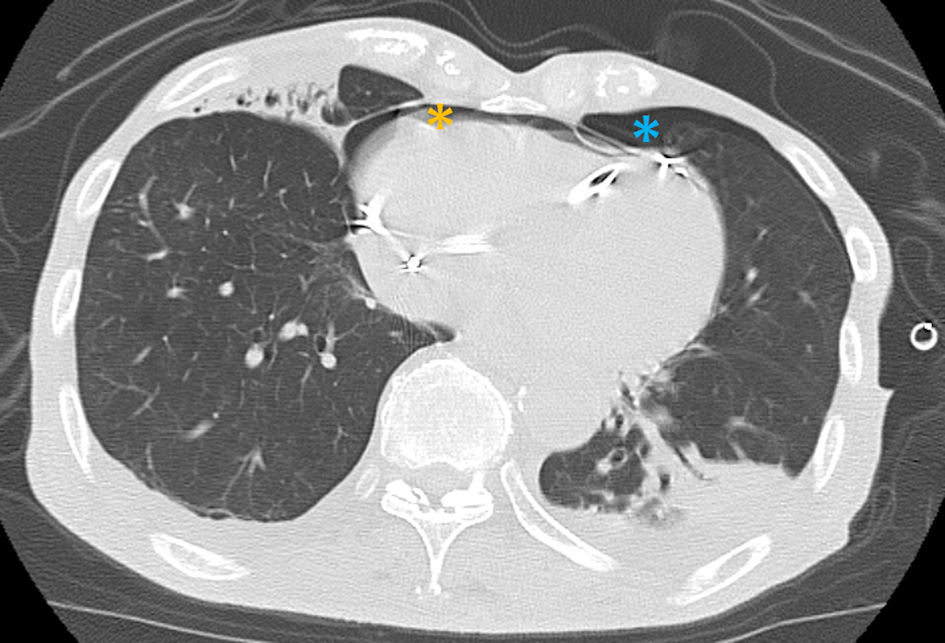
Figure 2. Right ventricular lead tip protruding into the left thoracic cavity ((a, c), white arrows), with moderate pneumopericardium and left severe pneumothorax, respectively ((b, c), orange and blue asterisks). (a) Scout image. (b) Axial image. (c) Coronal image.
Figure 3.
Figure 3. Both left pneumothorax (orange asterisk) and pneumopericardium (blue asterisk) were relieved 2 h after chest tube drainage.
Figure 4.
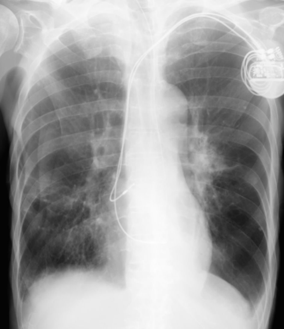
Figure 4. There was no recurrence of pneumothorax and pneumopericardium 1 month after repositioning of the right ventricular lead.
Figure 5.
Figure 5. The active-fixation ventricular lead sequentially perforates the right ventricular free wall, pericardium, left pleura and lung, resulting in left pneumothorax (a, b). Intrathoracic air then tracks into the pericardial cavity through the pericardium-pleural fistula (red arrow), resulting in pneumopericardium (c).