Cardiology Research, ISSN 1923-2829 print, 1923-2837 online, Open Access
Article copyright, the authors; Journal compilation copyright, Cardiol Res and Elmer Press Inc
Journal website https://cr.elmerpub.com

Original Article

Volume 000, Number 000, April 2025, pages 000-000


Impact of Systematic Use of Fractional Flow Reserve and Optical Coherence Tomography on Percutaneous Coronary Intervention Outcomes in Patients With Diabetes

Figures

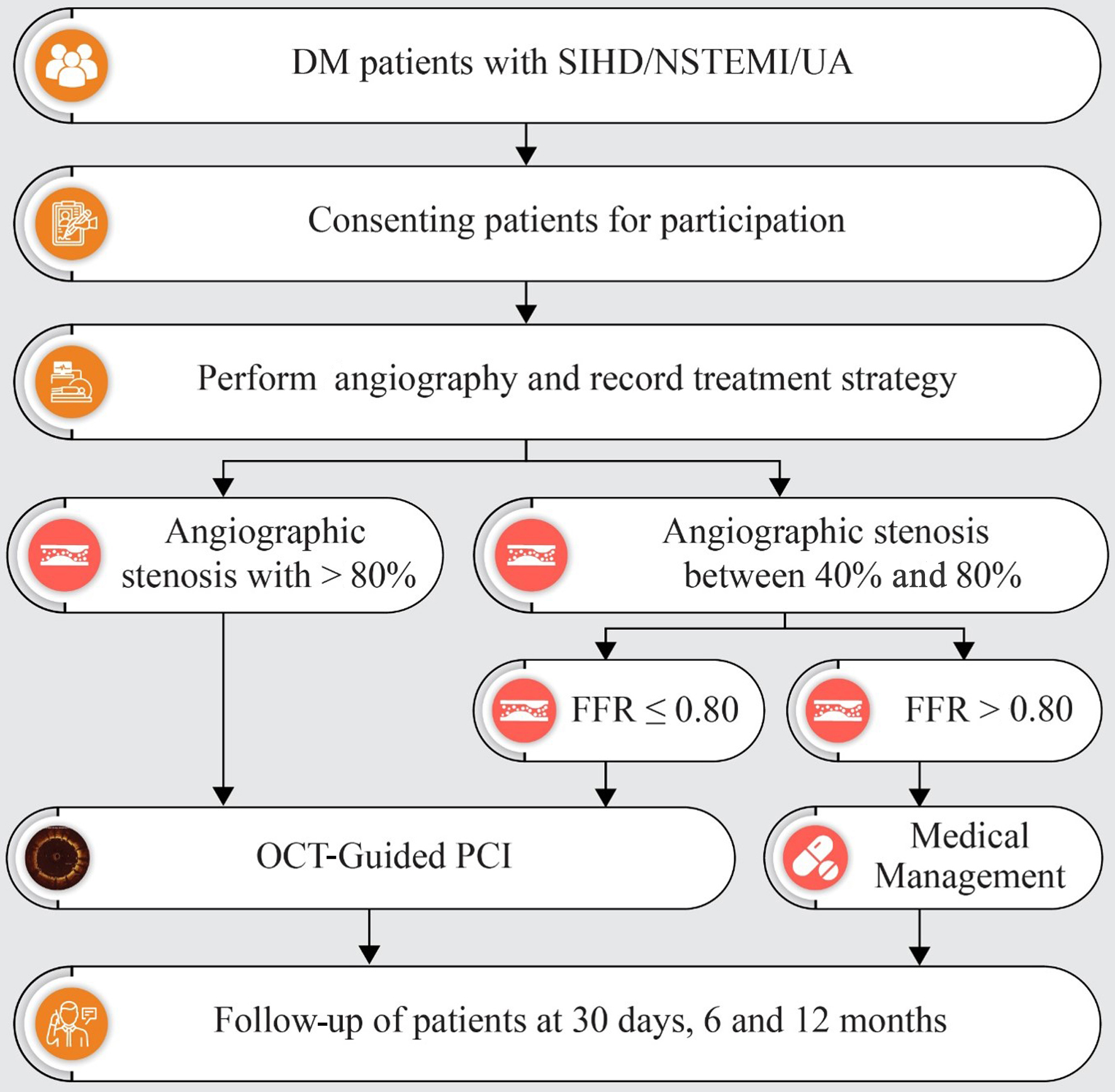
Figure 1.
Figure 1. Flowchart depicting the study plan and follow-up. DM: diabetes mellitus; FFR: fractional flow reserve; NSTEMI: non-ST-segment elevation myocardial infarction; OCT: optical coherence tomography; PCI: percutaneous coronary intervention; SIHD: stable ischemic heart disease; UA: unstable angina.
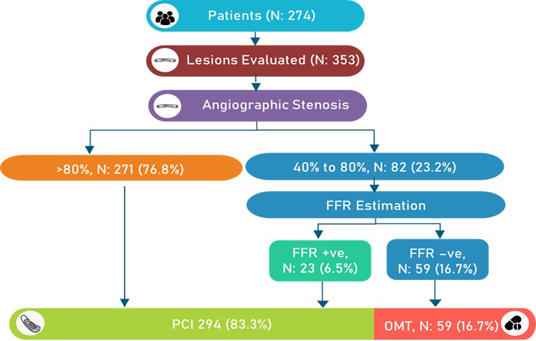
Figure 2.
Figure 2. Flowchart with the number of lesions and procedural details. FFR: fractional flow reserve; OMT: optimal medical therapy.
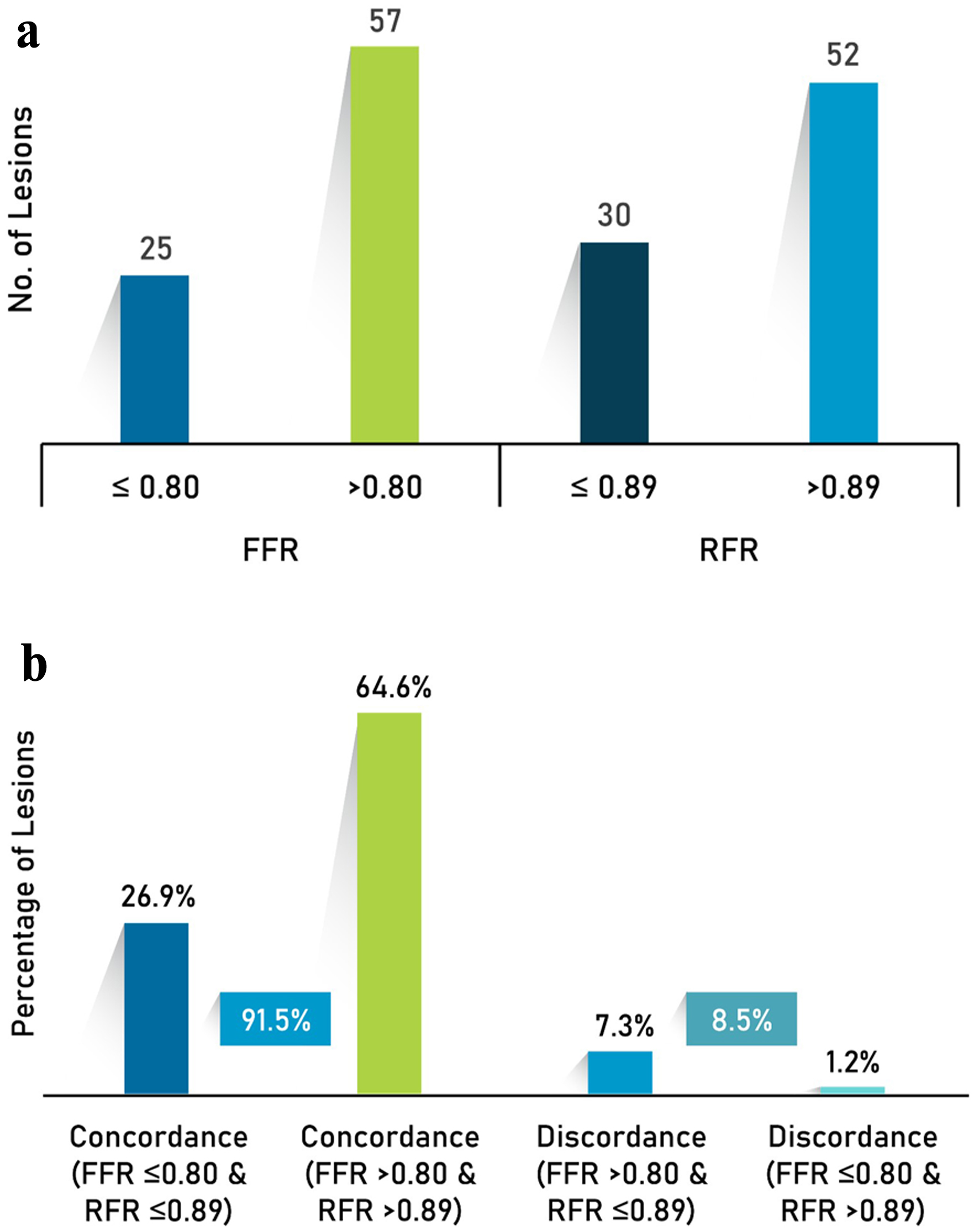
Figure 3.
Figure 3. (a) Physiology assessment and (b) concordance and discordance between RFR and FFR. FFR: fractional flow reserve; RFR: resting full-cycle ratio.
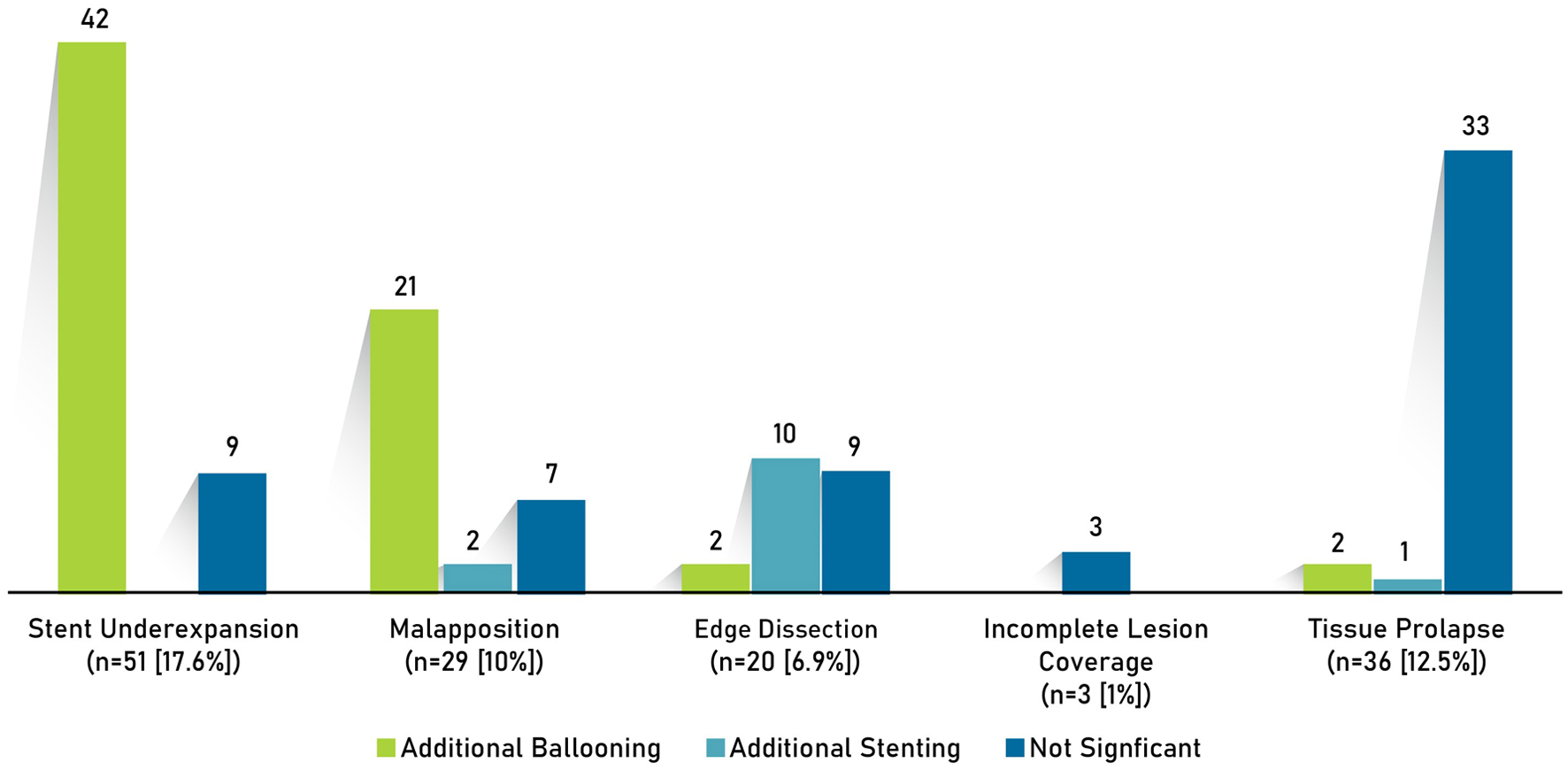
Figure 4.
Figure 4. Post-PCI optimization strategies. PCI: percutaneous coronary intervention.
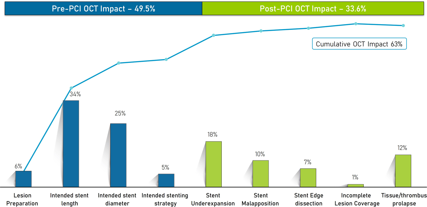
Figure 5.
Figure 5. Impact of OCT on pre- and post-PCI strategy. OCT: optical coherence tomography; PCI: percutaneous coronary intervention.

Tables

Table 1. Baseline Characteristics of the Study Population
 
CharacteristicsN = 274
BMI: body mass index; CAD: coronary artery disease; DPP4: dipeptidyl peptidase 4; FU: follow-up; GLP-1: glucagon-like peptide-1; MI: myocardial infarction; NSTEMI: non-ST-segment elevation myocardial infarction; PCI: percutaneous coronary intervention; SD: standard deviation; SGLT-2: sodium-glucose cotransporter 2.
Age, years, mean ± SD59.1 ± 9.4
BMI, kg/m226.2 ± 3.7
Male, n (%)203 (74.0)
Medical history, n (%)
  Systemic hypertension208 (75.9)
  Dyslipidemia62 (22.6)
  Smoking33 (12.0)
  Family history of CAD28 (10.2)
  Previous PCI26 (9.5)
  Previous MI17 (6.2)
  Pulmonary disease13 (4.7)
  Chronic kidney disease7 (2.6)
  Cerebral vascular accident2 (0.7)
Indication for PCI, n (%)
  Unstable angina119 (43.4)
  NSTEMI84 (30.7)
  Stable ischemic heart disease71 (25.9)
Duration of diabetes, months80.3 ± 78.5
Statins251 (91.6)
Antidiabetic medications, n (%)
  Insulin requiring111 (40.5)
  Biguanides (metformin)178 (65.0)
  DPP4 inhibitors (gliptins)63 (23.0)
  Sulfonylureas58 (21.2)
  SGLT-2 inhibitors (gliflozins)49 (17.9)
  Meglitinides (sulfonylurea analog)4 (1.5)
  GLP-1 receptor agonists (incretin)4 (1.5)
  Thiazolidinediones (glitazones)3 (1.1)
  Alpha-glucosidase inhibitors1 (0.4)
HbA1c (%)
  Baseline8.3 ± 1.9
  At 12 months FU7.8 ± 1.6
Serum creatinine (mg/dL)
  At baseline1.0 ± 0.2
  At discharge1.0 ± 0.4


Table 2. Lesion Description
 
CharacteristicsValue
ACC/AHA: American College of Cardiology/American Heart Association; D1: diagonal 1; LAD: left anterior descending; LCX: left circumflex; OM1: obtuse marginal 1; OM2: obtuse marginal 2; PDA: posterior descending artery; PLV: posterior left ventricular; RCA: right coronary artery; RI: ramus intermedius; SD: standard deviation.
Angiographic assessment
  SYNTAX score, mean ± SD12.1 ± 5.9
    Low (0 - 22), n (%)256 (93.4%)
    Intermediate (23 - 32), n (%)18 (6.6%)
  Small vessels (≤ 3 mm), n (%)218 (61.8%)
Disease disposition
  Multivessel and multiple lesions62 (22.6%)
  Multivessel and single lesions81 (29.6%)
  Single vessel and multiple lesions1 (0.4%)
  Single vessel and single lesions130 (47.4%)
Lesion types
  Bifurcation lesions, n (%)94 (26.6%)
  Long lesions (≥ 28 mm)219 (62.0%)
Lesion location, n (%)
  LAD, D1209 (59.2%)
  RCA, RI, PLV, PDA92 (26.1%)
  LCX, OM1, OM252 (14.7%)
Lesion complexity (ACC/AHA), n (%)
  A45 (12.7%)
  B1124 (35.1%)
  B267 (19%)
  C117 (33.1%)
Stenosis severity by angiography
  40-80%82 (23.2%)
  > 80%271 (76.8%)


Table 3. Lesion Assessment by Angiography vs. OCT (n = 289 Lesions)
 
AssessmentAngiographyOCTP-value
OCT: optical coherence tomography; RVD: reference vessel diameter; SD: standard deviation.
Need for pre-dilatation for stenting, n (%)257 (88.9)260 (90.0)0.6842
Need for lesions debulking, n (%)67 (23.2)85 (29.4)0.3924
  Cutting balloon27 (9.3)34 (11.8)
  Scoring balloon23 (7.9)39 (13.5)
  Intravascular lithotripsy11 (3.8)8 (2.8)
  Rotablation8 (2.8)8 (2.8)
Calcification, n (%)106 (36.7)138 (47.8)0.0804
Proximal RVD, mm (mean ± SD)3.2 ± 0.63.3 ± 0.60.0456
Distal RVD, mm (mean ± SD)2.8 ± 0.52.9 ± 0.60.0456
Lesion length, mm (mean ± SD)29.4 ± 13.832.0 ± 13.70.0234
Intended stent length, mm (mean ± SD)33.7 ± 14.635.5 ± 14.2< 0.0001
Intended stent diameter, mm (mean ± SD)2.9 ± 0.43.0 ± 0.50.0082
Intended number of stents, n (%)
  One250 (86.5)250 (86.5)-
  Two39 (13.5)39 (13.5)-