Figures
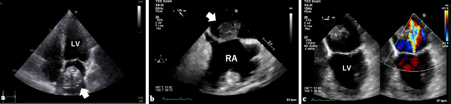
↓ Figure 1. Transthoracic (a) and transesophageal echocardiography (b) showed a large mass (white arrows) in a dilated left atrium. The mass was attached to the interatrial septum with a wide base. Moderate mitral regurgitation associated with annular dilatation was observed (c). LV: left ventricle; RA: right atrium.
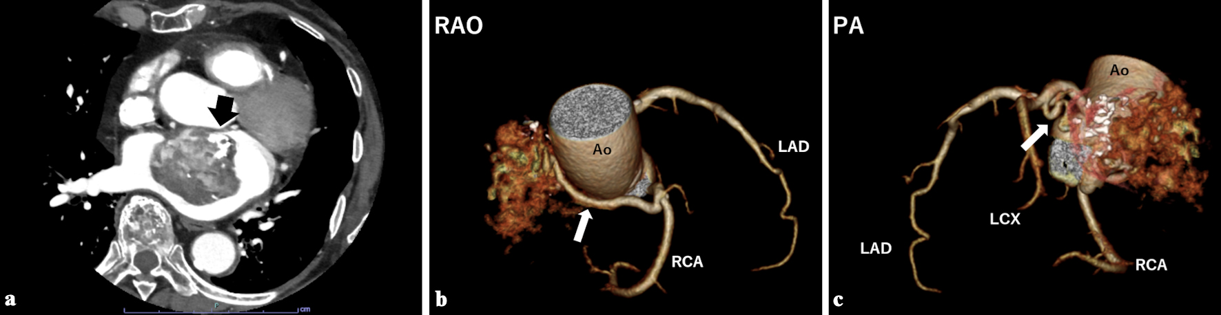
↓ Figure 2. (a) Contrast-enhanced chest computed tomography showed a heterogeneously enhanced mass with partial calcification (black arrow). Computed tomography coronary angiography showed two feeding vessels originating from the right coronary artery (b, white arrow) and the left circumflex artery (c, white arrow). Ao: aorta; LAD: left anterior descending artery; LCX: left circumflex artery; PA: posterior-anterior projection; RAO: right anterior oblique projection; RCA: right coronary artery.
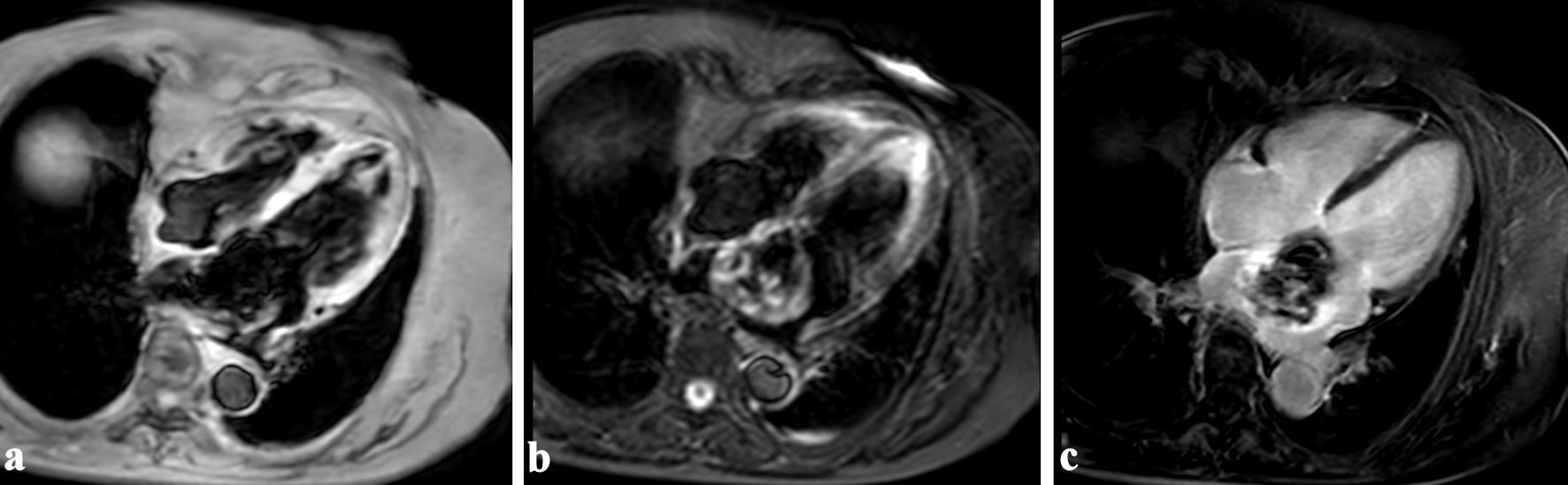
↓ Figure 3. Cardiac magnetic resonance imaging showed that the interior of the mass was hypointense on T1-weighted (a) and T2-weighted imaging (b), and the exterior was hyperintense on T2-weighted imaging. Heterogeneous enhancement was observed on late gadolinium enhancement imaging (c).
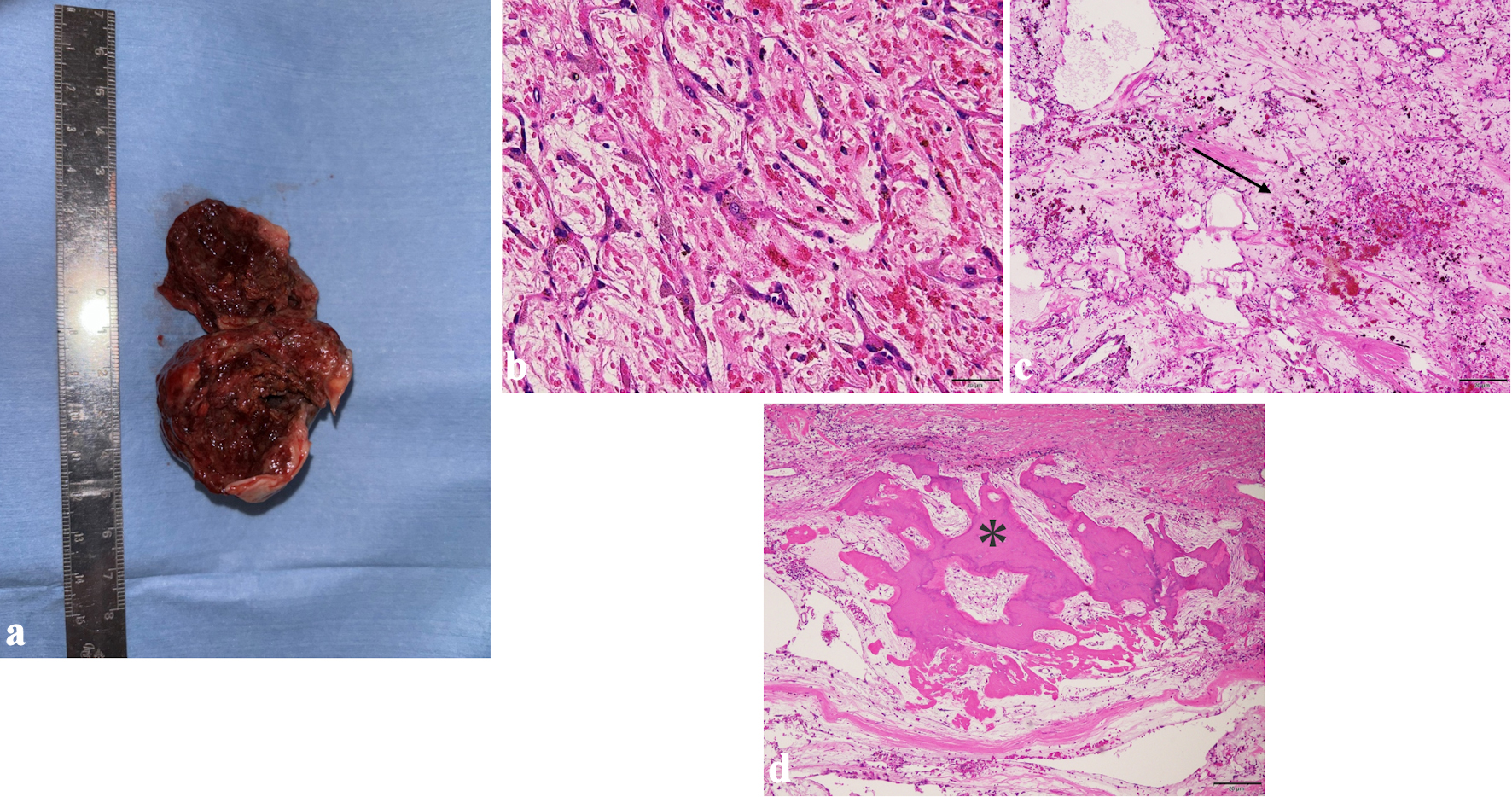
↓ Figure 4. The excised tumor was reddish brown and friable and measured 50 × 45 × 40 mm (a). Photomicrographs of hematoxylin and eosin (H&E) staining sections are shown: (b) H&E staining × 200; (c) H&E staining × 40; (d) H&E staining × 40. Proliferated cells with spindle-shaped nuclei were embedded in a myxoid matrix (b). The interior of the tumor contained hemorrhage (c, black arrow) and foci of ossification (d, asterisk).
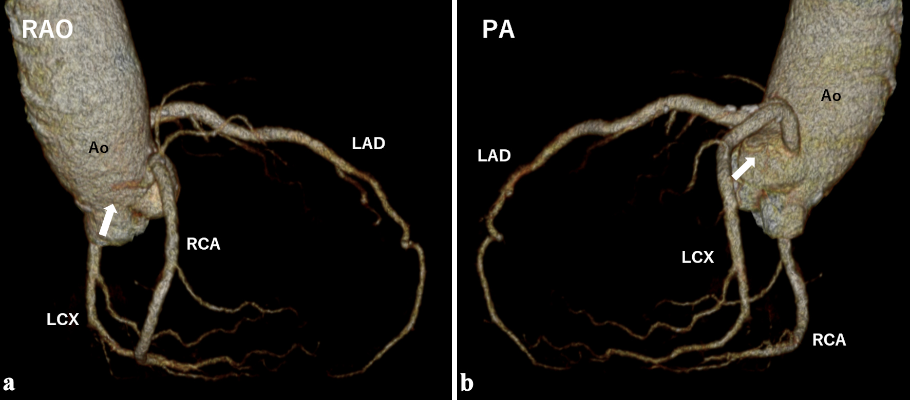
↓ Figure 5. Computed tomography coronary angiography performed 6 months postoperatively showed disappearance of the feeding vessels (white arrows). Ao: aorta; LAD: left anterior descending artery; LCX: left circumflex artery; PA: posterior-anterior projection; RAO: right anterior oblique projection; RCA: right coronary artery.