Figures
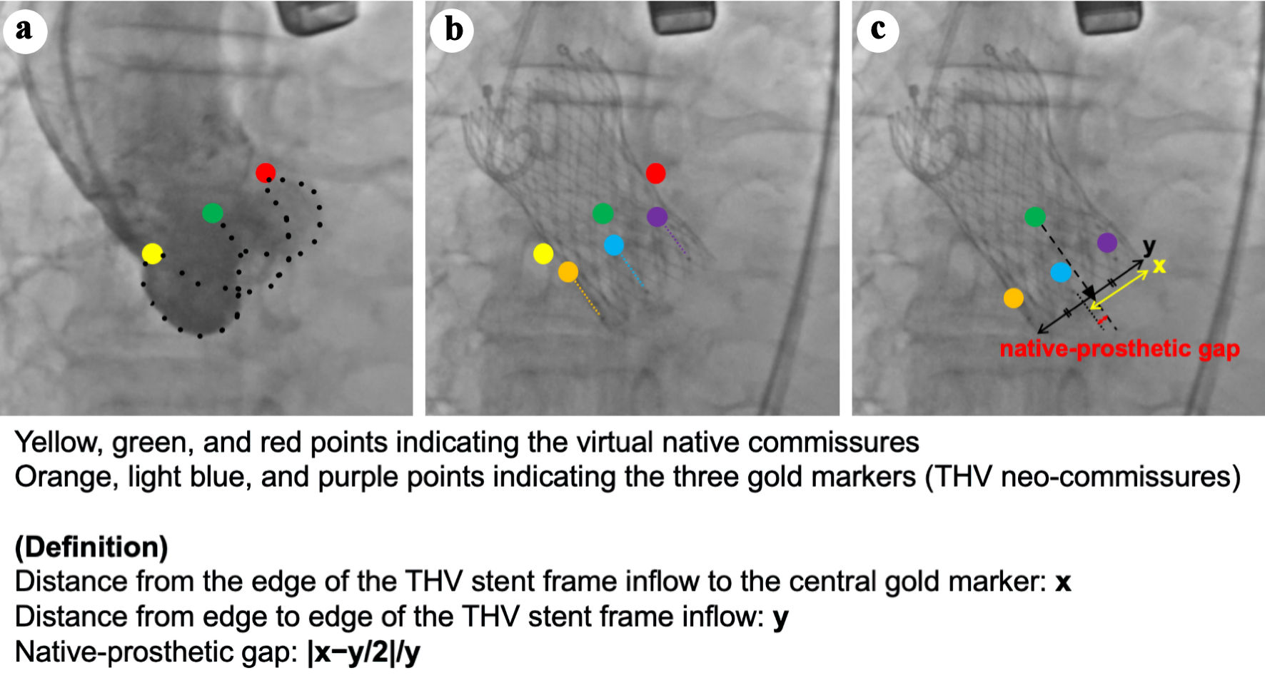
↓ Figure 1. Native-prosthetic gap (NPG). The method for calculating the NPG is as follows. (a) Initial aortography in the three-cusp coplanar view (3-CV) is shown, with yellow, green, and red points indicating the virtual native commissures. (b) The orientation of the gold markers in the 3-CV after transcatheter heart valve (THV) deployment. Orange, light blue, and purple points represent the three gold markers (i.e., THV neo-commissures). (c) The measurement of NPG. The yellow arrow indicates the distance between the end of the THV stent frame and the central gold marker (x), while the black arrow indicates the total length of the THV stent frame (y). The black triangle represents the center of the stent frame, with a distance of y/2 from the end of the THV stent frame. The NPG was calculated as |x-y/2|/y, representing the proportion of the distance between the center of the THV stent frame and the central gold marker relative to the total length of the THV stent frame. 3-CV: three-cusp coplanar view.
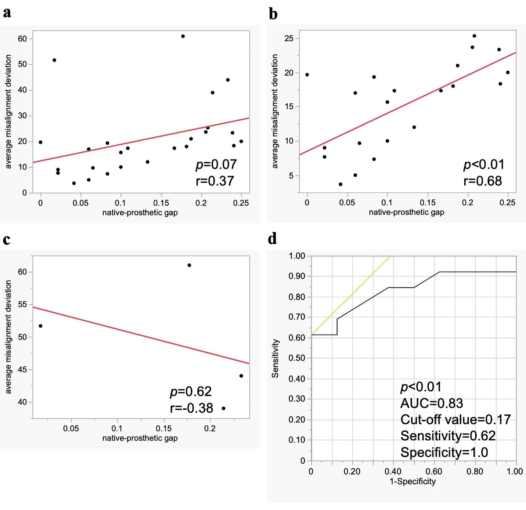
↓ Figure 2. Association between native-prosthetic gap and commissural misalignment (CMA). Linear regression analysis for CMA in all cases (a), in cases with commissural alignment and mild CMA (b), and in cases with moderate and severe CMA (c). Logistic regression and receiver operating characteristic analysis for CMA in cases with commissural alignment and mild CMA (d). AUC: area under the curve.
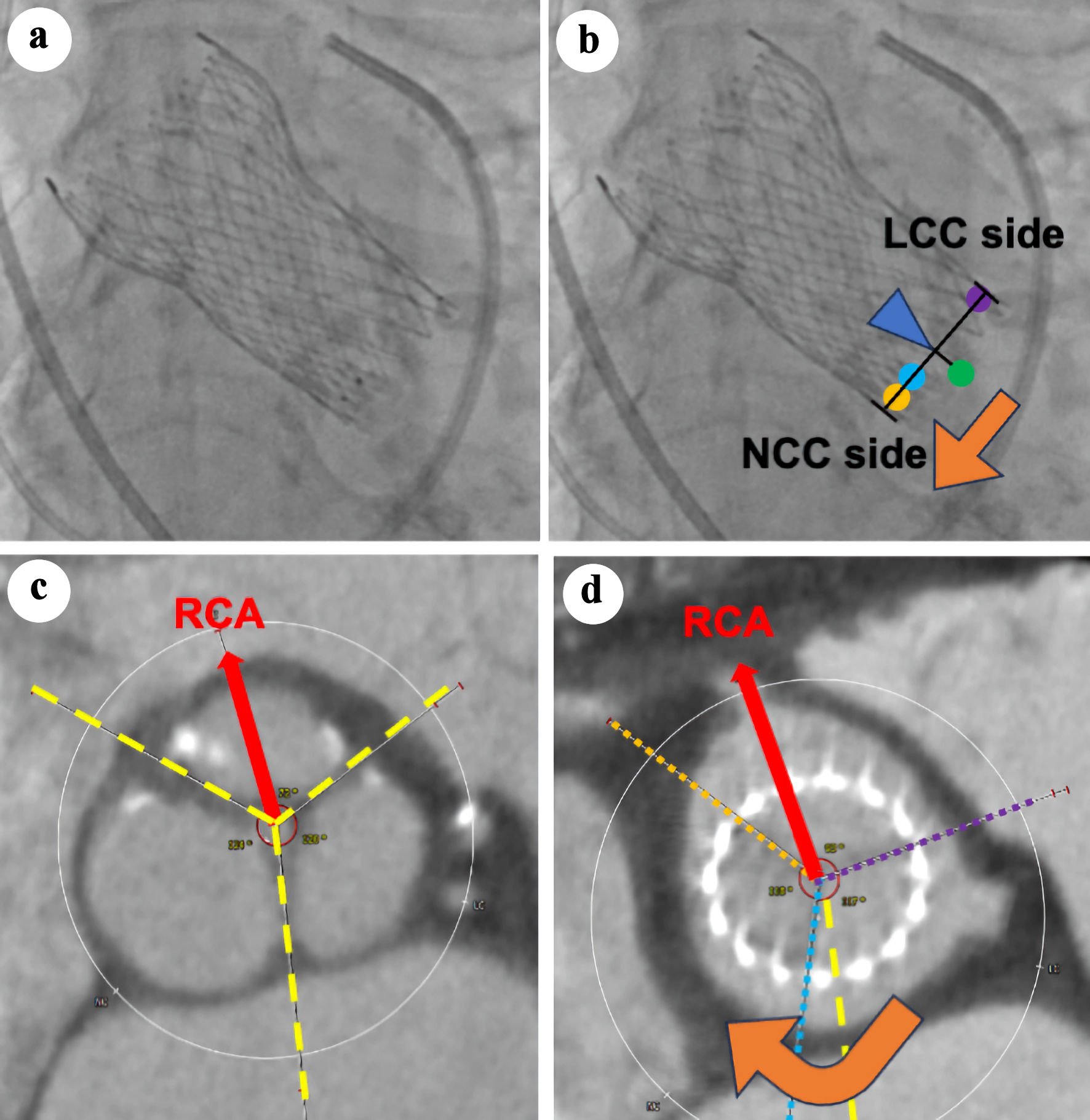
↓ Figure 3. Association between the direction of central marker shift in fluoroscopy and the direction of commissural misalignment in computed tomography (CT). (a, b) The transcatheter heart valve (THV) orientation images obtained in the three-cusp coplanar view on fluoroscopy after THV deployment. Orange, light blue, and purple points represent the three gold markers (i.e., THV neo-commissures). The green point represents the virtual native commissure, located at the center of the THV stent frame. (c, d) Preprocedural and postprocedural CT. The red arrow indicates the direction of the RCA. Yellow dashed lines mark the native commissures, while orange, light blue, and purple dotted lines denote the neo-commissures of the THV. Clockwise misalignment occurred when the central marker was positioned closer to the NCC side (orange arrow, (b, d)). NCC: non-coronary cusp; LCC: left coronary cusp; RCA: right coronary artery.
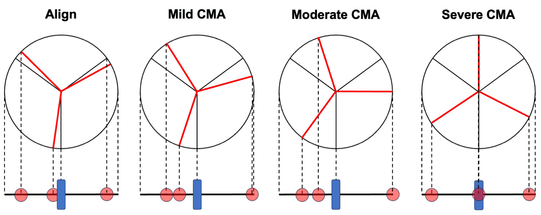
↓ Figure 4. Association between the orientation of the three gold markers and commissural misalignment (CMA). The relationship between CMA, as assessed by computed tomography, and the orientation of the gold markers (red circles) in fluoroscopy. As the degree of CMA progresses to mild, the central marker tends to move away from the center of the transcatheter heart valve (THV) stent frame (blue square). However, as it progresses to moderate and severe, the central marker paradoxically shifts closer to the center of the THV stent frame.
Tables
↓ Table 1. Baseline Clinical Characteristics
| All patients (n = 25) |
|---|
| Data are presented as the median (interquartile range) for continuous variables or the number (percent) of patients for categorical variables. NYHA: New York Heart Association functional classification; STS: Society of Thoracic Surgeons; TTE: transthoracic echocardiography; LVEF: left ventricular ejection fraction; AVA: aortic valve area; EOA: effective orifice area. |
| Age, years | 84 (80 - 89) |
| Male, n (%) | 9 (36%) |
| NYHA ≥ III, n (%) | 11 (44%) |
| STS score | 5.3 (3.3 - 9.6) |
| Coronary artery disease, n (%) | 2 (8%) |
| Preprocedural TTE | |
| LVEF, % | 67 (62 - 72) |
| Peak aortic velocity, m/s | 4.5 (4.3 - 5.5) |
| Mean aortic pressure gradient, mm Hg | 48 (42 - 72) |
| AVA, cm2 | 0.51 (0.35 - 0.78) |
| Aortic regurgitation | |
| Non or trivial, n (%) | 6 (24%) |
| Mild, n (%) | 14 (56%) |
| Moderate, n (%) | 5 (20%) |
| Postprocedural TTE | |
| LVEF, % | 68 (57 - 73) |
| Peak aortic velocity, m/s | 1.7 (1.2 - 2.2) |
| Mean aortic pressure gradient, mm Hg | 6 (3 - 10) |
| EOA, cm2 | 1.7 (1.4 - 2.4) |
| Aortic regurgitation | |
| None or trivial, n (%) | 12 (48%) |
| Mild, n (%) | 11 (44%) |
| Moderate, n (%) | 2 (8%) |
↓ Table 2. Procedure and Fluoroscopic Analysis
| THV: transcatheter heart valve; LAO: left anterior oblique; NPG: native-prosthetic gap. |
| THV size | |
| 23 mm, n (%) | 5 (20%) |
| 26 mm, n (%) | 9 (36%) |
| 29 mm, n (%) | 10 (40%) |
| 34 mm, n (%) | 1 (4%) |
| Implanting view | |
| Cusp overlap view, n (%) | 11 (44%) |
| Near-cusp overlap view, n (%) | 11 (44%) |
| LAO view, n (%) | 3 (12%) |
| 2 left-1 right in cusp overlap or near-cusp overlap view, n (%) | 17 (77%) |
| Final C-tab orientation | |
| Inner curve | 20 (80%) |
| Center front | 1 (4%) |
| Outer curve | 4 (16%) |
| Center back | 0 (0%) |
| NPG | 0.11 (0.06 - 0.21) |
↓ Table 3. CT Analysis
| CT: computed tomography; RCA: right coronary artery; RCC: right coronary cusp; LCC: left coronary cusp; NCC: non-coronary cusp; CMA: commissural misalignment. |
| Native annulus area, mm2 | 386 (316 - 462) |
| Native annulus perimeter, mm | 72 (65 - 77) |
| Pre RCA to RCC/LCC, ° | 75.0 (69.0 - 80.0) |
| Pre RCA to LCC/NCC, ° | 194.0 (181.0 - 203.0) |
| Pre RCA to NCC/RCC, ° | 317.0 (310.0 - 324.0) |
| Post RCA to RCC/LCC, ° | 87.0 (83.0 - 99.0) |
| Post RCA to LCC/NCC, ° | 210.0 (194.0 - 219.0) |
| Post RCA to NCC/RCC, ° | 327.0 (315.0 - 344.0) |
| Δ RCA to RCC/LCC, ° | 21.0 (9.0 - 31.0) |
| Δ RCA to LCC/NCC, ° | 17.0 (8.0 - 24.0) |
| Δ RCA to NCC/RCC, ° | 20.0 (9.0 - 28.0) |
| Average misalignment deviation, ° | 18.0 (10.0 - 24.0) |
| Commissural alignment category | |
| Commissural alignment, n (%) | 8 (32%) |
| Mild CMA, n (%) | 13 (52%) |
| Moderate CMA, n (%) | 2 (8%) |
| Severe CMA, n (%) | 2 (8%) |