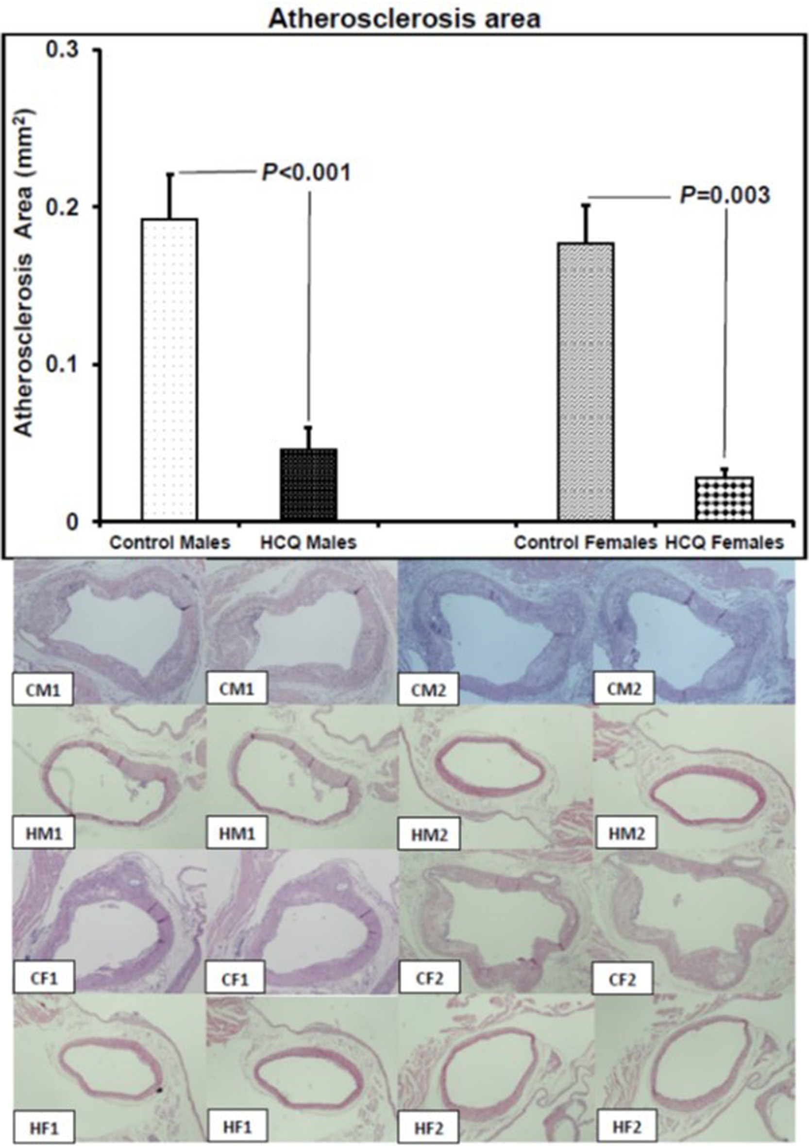
↓ Figure 1. Atherosclerosis area and representative aortic sections (hematoxylin and eosin stain, original magnification × 20) showing atherosclerotic lesions in two male and two female ApoE−/− control and hydroxychloroquine-treated (10 mg/kg/day) mice respectively, at 32 weeks of age, at the end of the 16-week treatment period. CM1: control male #1; CM2: control male #2; HM1: hydroxychloroquine-treated male #1; HM2: hydroxychloroquine-treated male #2; CF1: control female #1; CF2: control female #2; HF1: hydroxychloroquine-treated female #1; HF2: hydroxychloroquine-treated female #2. The left to right images are two representative histological sections, from the junction of the aorta to the heart and beyond into the ascending aorta up to the aortic arch, and used for histopathological evaluation of the entire lesion of each mouse. On histology, the atheromatous plaque of the control male and female mice is thicker than that of the hydroxychloroquine-treated male and female mice. All values are expressed as mean ± SEM. ApoE: apolipoprotein E.
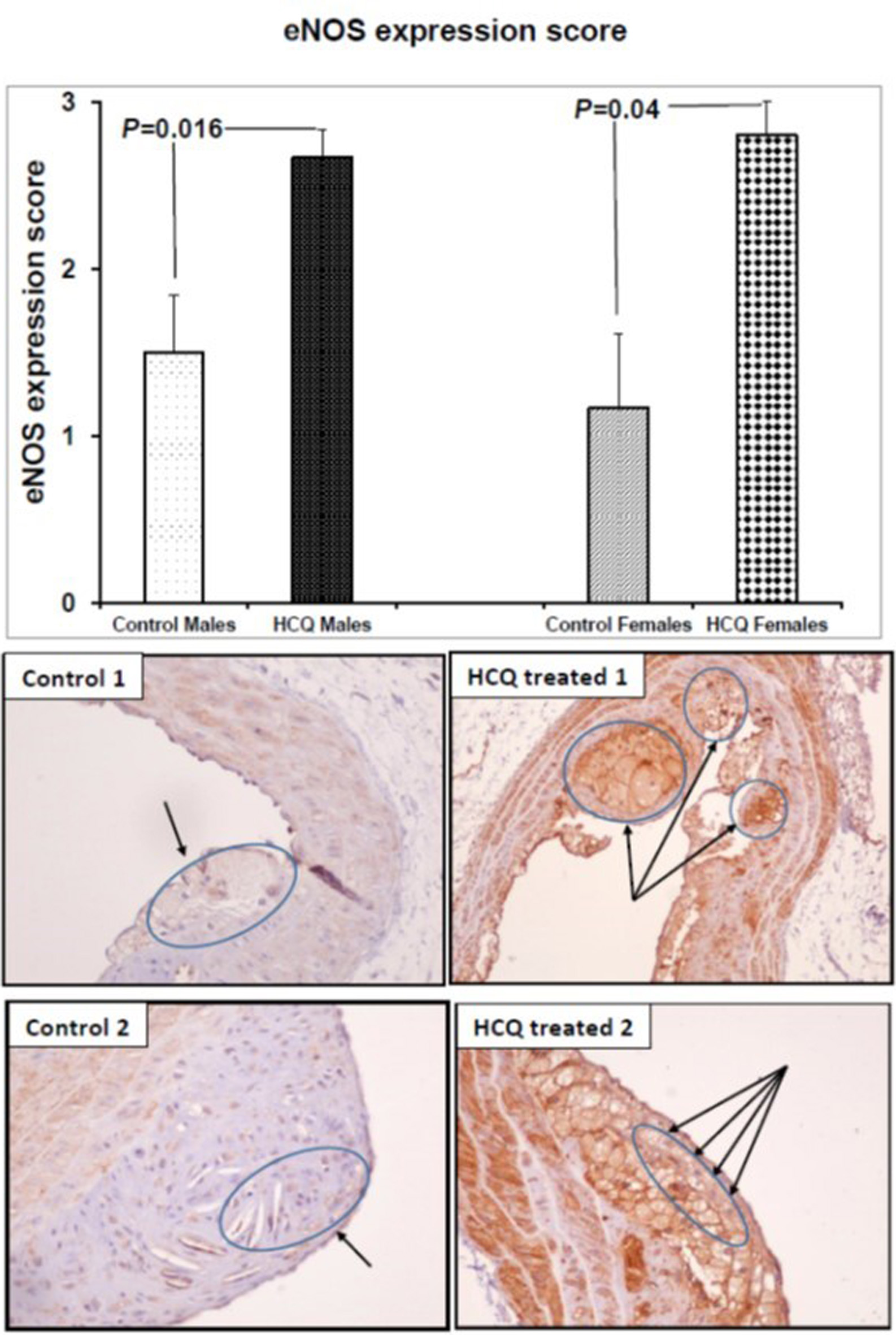
↓ Figure 2. eNOS expression score and representative eNOS immunostained aortic sections (× 20) in two ApoE−/− control and HCQ-treated (10 mg/kg/day) mice at 32 weeks of age, at the end of the 16-week treatment period. Immunohistochemically, the macrophages in the atheromatous plaque of the HCQ-treated mice show strong eNOS immunostaining (long arrows in right panel), whereas the immunoreaction is faint in the control mice plaque (short arrows in left panel). All values are expressed as mean ± SEM. ApoE: apolipoprotein E; eNOS: endothelial nitric oxide synthase; HCQ: hydroxychloroquine.
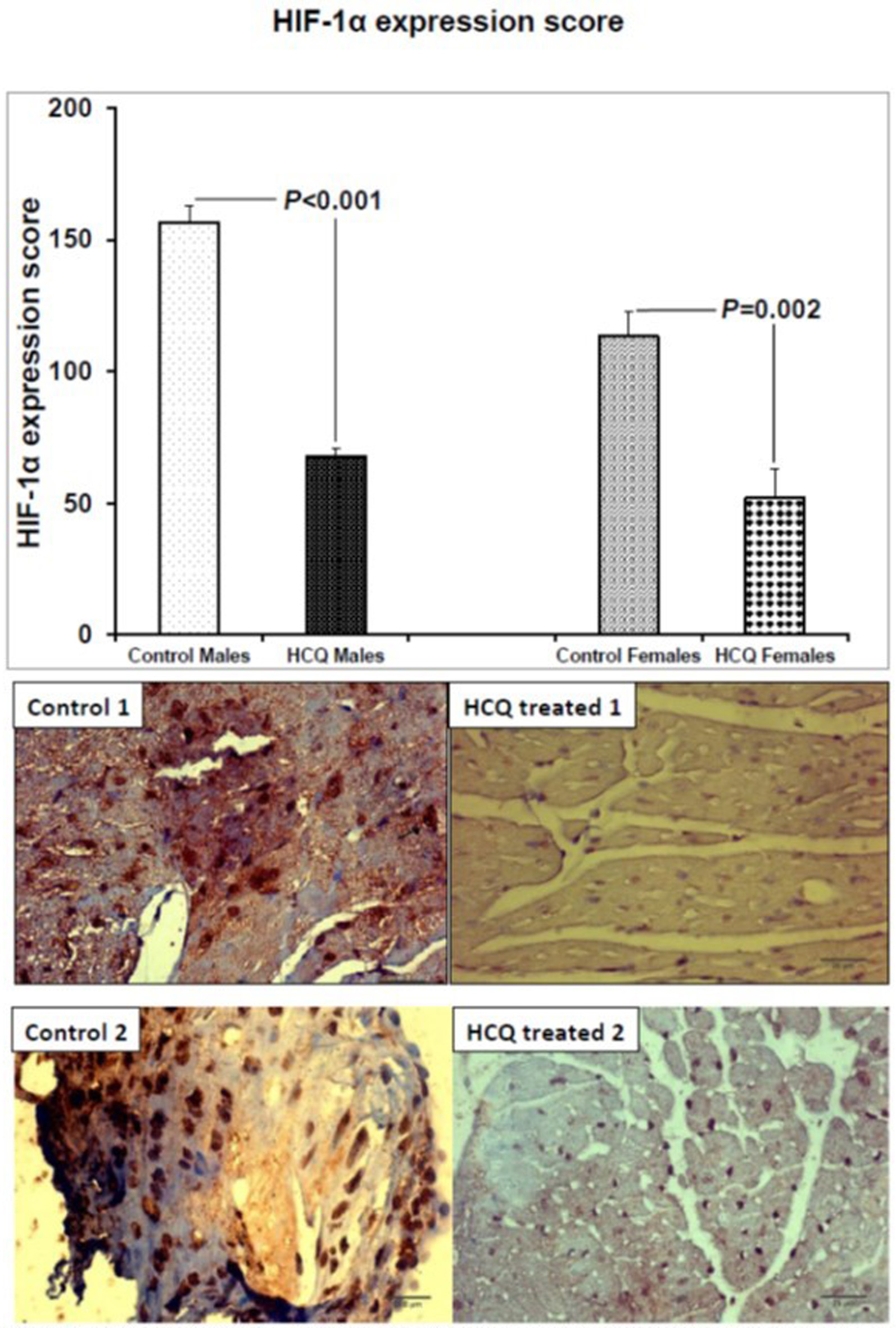
↓ Figure 3. HIF-1α expression score and representative HIF-1α immunostained aortic sections (×40) in two ApoE−/− control and HCQ-treated (10 mg/kg/day) mice at 32 weeks of age, at the end of the 16-week treatment period. Immunohistochemically, few macrophages in the atheromatous plaque of the HCQ-treated mice show weak nuclear HIF-1α immunostaining (blue nuclear staining in right panel), whereas the immunoreaction is strong in almost all nuclei of control mice atheromatous plaque (brown nuclear staining in left panel). All values are expressed as mean ± SEM. ApoE: apolipoprotein E; HCQ: hydroxychloroquine; HIF-1α: hypoxia-induced factor-1 alpha.