| Cardiology Research, ISSN 1923-2829 print, 1923-2837 online, Open Access |
| Article copyright, the authors; Journal compilation copyright, Cardiol Res and Elmer Press Inc |
| Journal website https://cr.elmerpub.com |
Review
Volume 17, Number 6, December 2026, pages 561-571

Clinically Actionable Prevention and Management of Major Complications During Durable Left Ventricular Assist Device Support: A Contemporary Narrative Review
Wei Yong Shenga, Tian Sheng Tanga, You Shi Wua, Hussain Asifa, Saifane Zakariaa, Da Fa Zhanga, b
aDepartment of Cardiovascular Surgery, The First Affiliated Hospital of Wannan Medical University (Yijishan Hospital), Wuhu, Anhui, China
bCorresponding author: Da Fa Zhang, Department of Cardiovascular Surgery, The First Affiliated Hospital of Wannan Medical University (Yijishan Hospital), Wuhu, Anhui, China
Manuscript submitted July 2, 2026, accepted August 28, 2026, published online October 24, 2026
Short title: LVAD Complication Management
doi: https://doi.org/10.14740/cr2283
| Abstract | ▴Top |
Durable left ventricular assist devices (LVADs) have become an established therapy for selected patients with advanced heart failure as bridge-to-transplant or destination therapy. Despite major progress in continuous-flow pump design and perioperative care, long-term benefit remains constrained by a recurrent burden of right heart failure, aortic regurgitation, pump thrombosis, stroke, bleeding, infection, and end-organ dysfunction. Because these events often arise from the interaction of altered hemodynamics, hemocompatibility-related injury, chronic antithrombotic exposure, and the practical complexity of longitudinal device care, a complication-centered management framework is clinically useful. This contemporary narrative review focuses on actionable prevention, surveillance, and treatment strategies for the major complications encountered during durable LVAD support. Rather than providing a broad device-oriented overview, we emphasize clinically relevant decision points across the perioperative and long-term course, including right ventricular protection, antithrombotic balance, structured monitoring for aortic regurgitation and hemolysis, prevention of driveline infection, and multidisciplinary outpatient follow-up. By organizing current evidence around practical management pathways, this review aims to support earlier recognition of adverse events and more durable long-term support.
Keywords: Durable left ventricular assist device; Advanced heart failure; Right heart failure; Hemocompatibility; Bleeding; Multidisciplinary follow-up
| Introduction | ▴Top |
Durable left ventricular assist devices (LVADs) are now an established form of mechanical circulatory support for selected patients with advanced heart failure, both as bridge-to-transplant and as destination therapy. Contemporary continuous-flow systems, particularly fully magnetically levitated centrifugal pumps, have improved device durability, hemocompatibility, survival, and quality of life compared with earlier-generation axial-flow pumps [1–3]. Design changes such as contact-free rotor suspension and wider blood-flow gaps are intended to reduce blood trauma, although clinical outcomes and management requirements remain device-specific [2, 3]. In our center, the practical experience informing this review is based primarily on HeartMate 3 and CorHeart 6 durable LVAD programs; therefore, the recommendations below are written for contemporary continuous-flow devices rather than for older pulsatile or axial-flow platforms. Device-specific trial findings should not be extrapolated between HeartMate 3 and CorHeart 6 without supporting evidence.
However, the major determinant of long-term success is no longer implantation alone. Instead, outcomes are increasingly shaped by the prevention, early recognition, and longitudinal management of major adverse events. Right heart failure (RHF), aortic regurgitation, pump thrombosis, stroke, bleeding, infection, and progressive end-organ dysfunction remain leading causes of rehospitalization, functional decline, and late mortality during LVAD support [2–5]. These complications rarely reflect isolated technical failures. More often, they emerge from the interaction of altered ventricular loading, continuous-flow physiology, hemocompatibility-related injury, chronic antithrombotic exposure, patient-device mismatch, and the operational demands of prolonged outpatient support.
An important challenge in the current literature is that complication domains are often discussed separately, whereas daily LVAD care requires integrated decisions across hemodynamics, anticoagulation, infection prevention, imaging surveillance, and multidisciplinary follow-up. For this reason, a clinically actionable synthesis should not simply catalogue complications; it should help clinicians recognize how these events evolve across the perioperative and chronic phases of support, where surveillance is most valuable, and when management thresholds should change.
This contemporary narrative review therefore focuses on the practical prevention, surveillance, and management of the major complications encountered during durable LVAD support. We specifically emphasize perioperative right ventricular (RV) protection, hemocompatibility-related adverse events, aortic valve surveillance, driveline infection prevention, end-organ monitoring, and multidisciplinary long-term care. Our aim is to provide a management-oriented framework that helps translate current evidence into day-to-day clinical practice rather than to present an exhaustive systematic review of all LVAD-related literature [5]. To make this framework operational, we summarize key complication domains, surveillance signals, and escalation thresholds in Table 1, provide a phase-based follow-up checklist in Table 2, and outline an integrated four-domain surveillance model in Figure 1. These tables are intended as a practical synthesis of contemporary evidence and expert clinical practice, rather than as validated risk algorithms or prescriptive institutional protocols.
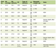 Click to view | Table 1. Practical Framework for Prevention, Surveillance, and Escalation of Major Complications During Durable LVAD Support |
Click to view | Table 2. Phase-Based Surveillance Checklist for Durable LVAD Recipients |
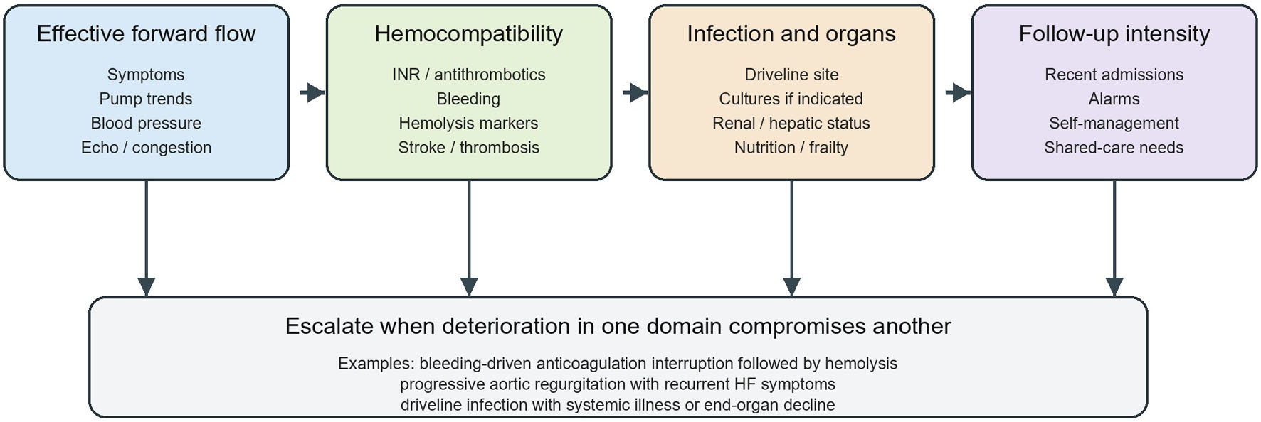 Click for large image | Figure 1. Four-domain surveillance and escalation framework for durable LVAD support. LVAD: left ventricular assist device. |
| Overview of Major Complications After LVAD Implantation | ▴Top |
Despite substantial improvement in outcomes after durable LVAD implantation, complications remain common and often determine whether support remains successful over the long term [2, 3, 6, 7]. For practical purposes, these complications can be grouped into hemodynamic complications, hemocompatibility-related adverse events, infectious complications, and end-organ dysfunction. Guideline- and consensus-based definitions are essential because similar symptoms may reflect different mechanisms. Post-LVAD RHF is generally defined by persistent or recurrent RV dysfunction requiring prolonged inotropes, inhaled or intravenous pulmonary vasodilators, right-sided mechanical circulatory support, or associated signs of venous congestion and end-organ dysfunction after LVAD implantation [4, 8]. Hemocompatibility-related adverse events include pump thrombosis, ischemic or hemorrhagic stroke, systemic embolism, hemolysis, and bleeding events requiring medical intervention, transfusion, hospitalization, or modification of antithrombotic therapy [2, 5–7, 9–14].
Hemodynamic complications mainly include RHF and progressive aortic regurgitation. These problems arise largely from altered ventricular interaction, RV preload and afterload imbalance, and chronic changes in valvular opening patterns during continuous-flow support [2, 4, 5, 8]. Hemocompatibility-related adverse events include pump thrombosis, systemic thromboembolism, hemolysis, and bleeding, particularly gastrointestinal bleeding. These events are closely related to non-physiological shear stress, platelet dysfunction, acquired von Willebrand syndrome, and the need for long-term antithrombotic therapy [2, 6, 7, 10, 11, 13].
Infectious complications, especially driveline infection and deeper device-related infection, remain a major source of late morbidity [15–18]. The 2024 International Society for Heart and Lung Transplantation (ISHLT) definitions distinguish mechanical circulatory support (MCS)-specific from non-MCS-specific infections [18]. Driveline infection should therefore be described by depth and extent rather than by erythema alone: superficial exit-site inflammation differs clinically from deep soft-tissue, tunnel, pump-pocket, or systemic infection. Renal dysfunction is also common during durable LVAD support and may reflect the combined effects of pre-existing heart failure, venous congestion, perioperative injury, and RV dysfunction [19]. Other end-organ complications and LVAD-related emergencies further complicate long-term care and often contribute to recurrent hospitalization [20].
Taken together, these complications reflect how device design, perioperative care, anticoagulation strategy, surveillance, and multidisciplinary management all shape long-term outcomes.
Routine follow-up can be organized around four recurring domains: (1) effective forward flow, assessed by symptoms, pump trends, blood pressure, echocardiography, and congestion; (2) hemocompatibility balance, assessed by anticoagulation stability, bleeding, hemolysis markers, and thromboembolic events; (3) infection and end-organ status, assessed by driveline findings, cultures when indicated, renal/hepatic function, nutrition, and frailty; and (4) follow-up intensity, adjusted according to recent admissions, alarms, recurrent adverse events, and patient self-management capacity. Escalation is appropriate when deterioration in one domain begins to compromise another, such as bleeding-driven anticoagulation interruption followed by hemolysis, progressive aortic regurgitation (AR) with recurrent heart failure symptoms, or driveline infection with systemic illness.
| RHF: Risk Stratification and Perioperative Management | ▴Top |
Pathophysiological mechanisms
RHF remains one of the defining and most difficult complications after LVAD implantation. RHF is multifactorial and may involve impaired RV-pulmonary vascular coupling under the new loading conditions created by left ventricular (LV) unloading. LVAD activation increases systemic flow and venous return, while leftward septal shift may impair RV geometry, tricuspid leaflet coaptation, and contractile efficiency. If pulmonary vascular resistance remains elevated, the RV faces simultaneous preload stress, afterload mismatch, septal dependence, and perioperative inflammatory or ischemic injury [4, 8, 21, 22].
Perioperative factors such as cardiopulmonary bypass, ischemia-reperfusion injury, volume shifts, bleeding, and inflammatory activation may further impair RV performance. Late RHF may also develop during chronic support and may be associated with persistent pulmonary vascular disease, progressive valvular dysfunction, arrhythmias, or suboptimal LVAD speed settings [4, 5, 8].
Preoperative predictors and risk stratification
Accurate prediction of RHF after LVAD implantation remains difficult, but risk stratification should be structured rather than impressionistic. Preoperative assessment should integrate echocardiography, right heart catheterization, laboratory evidence of congestion, and clinical trajectory. Useful echocardiographic variables include RV size, qualitative RV systolic function, tricuspid regurgitation severity, tricuspid annular plane systolic excursion (TAPSE), tissue Doppler S', fractional area change, RV free-wall strain when available, septal position, and inferior vena cava congestion. Hemodynamic variables include right atrial pressure, pulmonary artery pressure, pulmonary vascular resistance, pulmonary capillary wedge pressure, cardiac index, right atrial pressure/pulmonary capillary wedge pressure ratio, pulmonary artery pulsatility index, and RV stroke work index [4, 8, 21, 22].
Several risk scores have been proposed, including the Michigan Right Ventricular Failure Risk Score, CRITT score, EUROMACS-RHF score, and other models incorporating renal dysfunction, hepatic dysfunction, vasopressor requirement, right atrial pressure, Interagency Registry for Mechanically Assisted Circulatory Support (INTERMACS) profile, and RV dysfunction. The EUROMACS-RHF score, for example, incorporates INTERMACS class, use of multiple inotropes, severe RV dysfunction on echocardiography, right atrial pressure/pulmonary capillary wedge pressure ratio, and hemoglobin, with reported early severe RHF risk ranging from approximately 11% in low-risk patients to more than 40% in high-risk patients [8]. These scores can support preoperative discussion and planning, but none should replace invasive hemodynamic assessment and multidisciplinary judgment because performance varies across devices, eras, and institutional practices [4, 8, 21].
Prevention strategies
Prevention of RHF begins before surgery. Optimization of volume status means active decongestion until systemic venous congestion improves, including lower right atrial pressure, improved renal and hepatic indices, reduced peripheral edema or ascites, and acceptable filling pressures rather than simply achieving a lower body weight. Reduction of pulmonary vascular resistance may include oxygenation, correction of acidosis and hypercapnia, treatment of sleep-disordered breathing or pulmonary disease when relevant, cautious use of inotropes, and selective pulmonary vasodilators such as inhaled nitric oxide, inhaled prostacyclin, or phosphodiesterase-5 inhibition in appropriate patients [4, 8]. Careful patient selection and preoperative hemodynamic stabilization may reduce the likelihood of perioperative RV decompensation.
Intraoperatively, efforts should focus on minimizing RV ischemic injury, avoiding excessive transfusion, preventing hypoxia, hypercapnia, acidosis, and high airway pressures, and preventing abrupt septal displacement caused by aggressive pump speed settings. Transesophageal echocardiography should be used to assess RV size and function, septal position, tricuspid regurgitation, LV decompression, inflow cannula orientation, aortic valve opening, and adequacy of preload. Early postoperative surveillance should combine echocardiography, invasive hemodynamics when needed, renal and hepatic function, lactate, urine output, and serial congestion assessment [4, 8]. Prompt recognition of RV dysfunction may allow intervention before progression to refractory hemodynamic failure [4].
Management
Management of RHF after LVAD implantation includes decongestion, inotropes, pulmonary vasodilators, rhythm control, ventilatory optimization, and careful pump-speed adjustment. Pump speed should be titrated to maintain effective LV unloading without excessive leftward septal shift, suction events, worsening tricuspid regurgitation, or collapse of LV dimensions. In stable patients, echocardiography-guided ramp testing can help evaluate LV size, septal position, aortic valve opening, mitral regurgitation, RV response, estimated filling pressures, and pump parameters across speed settings. In unstable perioperative patients, intraoperative or bedside echo-guided speed adjustment is often more practical than formal ramp testing. In selected patients with severe or refractory RHF, temporary RV mechanical support should be considered early rather than after irreversible renal, hepatic, or metabolic deterioration [4, 8, 21, 22].
In practice, RHF is easier to manage when approached as a perioperative syndrome that requires planned prevention, not rescue treatment alone.
| AR: Mechanisms, Surveillance, and Intervention | ▴Top |
Mechanisms
AR is recognized with increasing frequency during chronic LVAD support, particularly when continuous-flow support is prolonged. Persistent reduction in native transvalvular pulsatility may contribute to incomplete aortic valve opening, commissural fusion, leaflet remodeling, and progressive valvular incompetence. Altered aortic root flow patterns may also contribute to de novo or worsening AR [23, 24].
Clinical impact and surveillance
Progressive AR may create a blind loop in which blood recirculates from the aorta into the LV and back through the pump, thereby reducing effective systemic output despite apparently acceptable device flow. Clinically, this may present with recurrent heart failure symptoms, reduced exercise tolerance, higher filling pressures, and repeated hospitalization [23, 24]. Echocardiography remains central to surveillance, but should be interpreted alongside pump parameters and overall clinical hemodynamics [23, 24].
Management
Management depends on severity, symptoms, mechanism, and surgical candidacy. Mild AR may initially be managed conservatively through blood pressure control and pump optimization, with attention to maintaining intermittent aortic valve opening when feasible. More advanced disease may require surgical aortic valve repair, closure, replacement, redo surgical aortic valve replacement, or transcatheter aortic valve replacement in selected high-risk patients [23–25]. In LVAD-supported patients, TAVR can be technically challenging because of annular dilation, limited calcification, altered root flow, and device-related hemodynamics, but it may provide a less invasive option for severe symptomatic AR in patients at prohibitive surgical risk. Redo SAVR or valve closure may provide more definitive correction in selected patients but carries substantial operative risk. Aortic root thrombosis or aortic valve thrombus should also be considered when the aortic valve remains persistently closed, particularly in the setting of low pulsatility, inadequate anticoagulation, or embolic events [23–25]. In practice, the central question is whether AR has become hemodynamically significant enough to compromise effective support [23, 24, 26–29].
| Pump Thrombosis and Thromboembolic Events: Optimizing Antithrombotic Balance | ▴Top |
Pathophysiology and risk factors
Pump thrombosis and thromboembolic complications remain major concerns despite substantial improvements in device design and hemocompatibility with contemporary continuous-flow LVADs. Their development is multifactorial and reflects the interplay of altered flow dynamics, blood-material interaction, incomplete endothelial adaptation, patient-specific prothrombotic tendency, subtherapeutic anticoagulation, infection, and device-related factors such as inflow or outflow malposition [2, 9, 11–14]. In the HeartMate 3 era, suspected or confirmed pump thrombosis is uncommon compared with earlier axial-flow devices. In the 5-year MOMENTUM 3 follow-up, device thrombosis was uncommon with the fully magnetically levitated centrifugal pump, compared with a substantially higher rate in the axial-flow comparator group, while stroke and bleeding remained clinically important residual risks [3]. Registry reports similarly show that hemocompatibility-related adverse events have decreased but not disappeared [6, 7, 11].
Clinically, thrombosis in LVAD recipients rarely behaves like an isolated mechanical event. It often develops in the setting of broader hemocompatibility disturbance. Hemolysis, inflammatory activation, low-grade flow disturbance, or concurrent infection may all precede overt thrombosis. Likewise, strategies that reduce bleeding risk too aggressively may increase thrombotic vulnerability in selected patients.
Clinical presentation and diagnostic evaluation
The presentation of pump thrombosis is often heterogeneous. Patients may develop progressive heart failure symptoms, low-flow alarms, increased pump power consumption, dark urine, laboratory evidence of hemolysis, or recurrent embolic events [2, 11, 12, 27]. Lactate dehydrogenase remains one of the most practical biomarkers in clinical monitoring, although interpretation should be integrated with plasma-free hemoglobin, pump parameters, symptoms, and imaging findings [12].
Echocardiography and device log review remain central to evaluation, while computed tomography (CT) may help define cannula position or outflow graft abnormalities. Because delayed recognition can lead to refractory hemolysis, stroke, or urgent reoperation, diagnostic escalation should not be postponed when clinical suspicion is high [20, 27].
Prevention and treatment strategies
The prevention of pump thrombosis depends largely on maintaining antithrombotic balance. During implantation, unfractionated heparin is commonly administered before cardiopulmonary bypass with an activated clotting time (ACT) target determined by institutional cardiopulmonary bypass protocols. After surgery, intravenous heparin is usually restarted only after surgical bleeding is controlled, chest tube output is acceptable, and coagulation status is stable; many centers begin low-dose heparin within the first 12–24 h and transition toward therapeutic anticoagulation over the following days, but timing must be individualized [5, 9]. Long-term therapy has traditionally used warfarin with a device-specific international normalized ratio (INR) target and aspirin, although the need for aspirin is being reconsidered in HeartMate 3 recipients after the ARIES-HM3 trial showed that aspirin avoidance reduced nonsurgical bleeding without increasing thrombotic events in appropriately selected patients [30]. Direct oral anticoagulants should not be used routinely for durable LVAD anticoagulation outside clinical trials or highly selected specialist-directed circumstances. Prior experience with dabigatran in mechanical circulatory support raised safety concerns, and evidence for factor Xa inhibitors remains insufficient for broad recommendation. Therefore, vitamin K antagonist-based anticoagulation remains the standard backbone for most durable LVAD recipients, with antiplatelet therapy individualized by device type, bleeding history, thrombotic risk, and institutional protocol [5, 30].
When pump thrombosis is suspected or confirmed, treatment options include intensification of anticoagulation, short-term parenteral therapy, thrombolysis in carefully selected patients, or surgical pump exchange when conservative treatment is ineffective or unsafe [5, 20]. The choice depends on hemodynamic status, neurological risk, bleeding risk, and the severity of device dysfunction.
Stroke and systemic thromboembolism
Stroke remains one of the most serious complications during LVAD support because of its major impact on mortality, disability, and subsequent treatment options. Risk is shaped by antithrombotic control, device-related thrombogenicity, blood pressure management, infection, and patient comorbidity [2, 11, 14, 31]. Contemporary registry and trial data suggest that stroke remains a clinically meaningful event even with magnetically levitated devices; stroke remains a concern during long-term HeartMate 3 support, although rates vary according to device era, follow-up duration, event definitions, blood pressure control, infection burden, and antithrombotic strategy [2, 3, 6, 7, 14, 31]. Prevention therefore requires integrated management, including stable anticoagulation, infection control, blood pressure optimization, and early recognition of hemolysis or device dysfunction [5, 14, 31].
| Bleeding Complications: Hemocompatibility-Related Injury and Clinical Management | ▴Top |
Mechanisms of bleeding in continuous-flow LVAD support
Bleeding is among the most frequent complications following LVAD implantation and remains a major cause of recurrent hospitalization [2, 6, 13]. Its pathophysiology extends beyond simple over-anticoagulation. Continuous-flow devices expose blood components to non-physiological shear stress, contributing to platelet dysfunction, degradation of high-molecular-weight von Willebrand factor multimers, and acquired von Willebrand syndrome. Prior mechanistic and clinical discussions suggest that von Willebrand factor degradation is an important link between shear stress, hemolysis, and bleeding during LVAD support. Reduced pulsatility may also promote gastrointestinal angiodysplasia, particularly during long-term device support [13].
The clinical result is a fragile hemostatic balance in which even otherwise standard antithrombotic regimens may provoke recurrent bleeding. This explains why bleeding remains common even in patients with apparently therapeutic anticoagulation levels and why management often requires repeated reassessment rather than one-time correction.
Gastrointestinal bleeding
Gastrointestinal bleeding is the most characteristic bleeding phenotype during durable LVAD support. It may present as overt upper or lower gastrointestinal hemorrhage, recurrent occult blood loss with anemia, or repeated hospital admissions for transfusion and endoscopic evaluation. The recurrence rate is clinically significant and often contributes to treatment fatigue, impaired quality of life, and repeated interruption of anticoagulation [6, 7, 13].
The pathogenesis is multifactorial, involving mucosal hypoperfusion, vascular malformation, altered angiogenic signaling, acquired von Willebrand syndrome, and chronic anticoagulant exposure. Because of this complexity, endoscopic management alone may not fully prevent recurrence [13].
Management principles
Management of bleeding should begin with hemodynamic stabilization and risk stratification. Temporary adjustment of anticoagulation or antiplatelet therapy may be required, but this must be balanced carefully against the competing risk of pump thrombosis or stroke [5, 10, 11, 13, 14]. Endoscopic evaluation is appropriate for gastrointestinal bleeding when clinically indicated, and transfusion strategies should be individualized according to hemodynamic status and comorbidity.
In patients with recurrent bleeding, management often shifts from treatment of isolated episodes to long-term recurrence prevention. This may include reassessment of antithrombotic intensity, optimization of blood pressure, evaluation of device parameters, and in selected cases a broader reassessment of overall support strategy [5–7, 13, 30].
Prevention strategies
Prevention of bleeding in LVAD recipients requires more than conservative anticoagulation alone. It also depends on minimizing unnecessary antithrombotic exposure, monitoring for recurrent anemia or occult bleeding, and recognizing patients in whom a lower-intensity strategy may be reasonable. In the contemporary era, there is growing interest in individualized regimens, particularly in HeartMate 3 recipients, but careful monitoring remains essential [5, 10, 13, 30].
| Infection and Driveline-Related Complications | ▴Top |
Clinical spectrum
Infectious complications remain a persistent limitation of long-term LVAD support. Driveline infections are the most common device-related infections, although deeper pump or pocket infections and bloodstream infections also occur. These events can lead to recurrent hospitalization, compromise transplant candidacy, and adversely affect long-term survival [5, 18, 32].
Risk factors and diagnosis
Multiple factors contribute to infection risk, including diabetes, malnutrition, renal dysfunction, prolonged support duration, inadequate exit-site care, local trauma, and repeated dressing disruption. Diagnosis requires careful attention to the depth and extent of infection. Superficial exit-site inflammation must be distinguished from deeper tissue involvement, tunnel infection, or systemic infection, as management strategies differ substantially.
Clinical examination, wound assessment, microbiological cultures, inflammatory markers, and selected imaging studies all contribute to diagnosis. Recommended surveillance should include exit-site pain, erythema diameter, warmth, swelling, induration, drainage amount and character, granulation tissue, driveline trauma or instability, fever, leukocytosis, C-reactive protein when clinically indicated, blood cultures for systemic illness, and ultrasound or CT/positron emission tomography (PET)-CT when deep extension is suspected [15–18]. Recurrent or indolent symptoms should be taken seriously, particularly in patients on chronic support.
Treatment and prevention
Treatment generally includes targeted antimicrobial therapy, local wound care, and, in some cases, surgical intervention. In selected patients who are not immediate candidates for device exchange or transplantation, chronic suppressive treatment may also be necessary. Prevention is the more important long-term strategy and depends heavily on patient education, driveline stabilization, standardized dressing protocols, and regular outpatient surveillance [5, 18, 32, 33]. Recent quality-improvement work has emphasized structured infection-reduction toolkits and standardized preventive workflows across centers, suggesting that infection prevention should be built into routine care rather than left to local improvisation [33].
Recent prospective work has explored additional preventive approaches, including local prophylactic strategies and alternative driveline positioning techniques, but the evidence remains limited [16, 17].
Further progress may come from fully implantable or wirelessly powered systems that reduce long-term reliance on percutaneous hardware.
| Renal Dysfunction and Other End-Organ Complications | ▴Top |
Renal dysfunction is common both before and after LVAD implantation and reflects the combined effects of advanced heart failure, venous congestion, low cardiac output, perioperative injury, and RV dysfunction [19]. Some patients improve after LV unloading, whereas others develop persistent or progressive renal impairment, particularly when postoperative congestion, RHF, or repeated complications persist [5, 19].
Renal function has direct implications for anticoagulation safety, infection risk, diuretic responsiveness, and transplant candidacy. For that reason, it deserves longitudinal follow-up alongside hemodynamic assessment rather than being treated as a secondary comorbidity.
Other end-organ complications, including hepatic dysfunction, malnutrition, frailty progression, and arrhythmias, may likewise reflect the overall adequacy of support and the cumulative burden of adverse events [5, 19, 20]. These problems often interact and contribute to recurrent hospitalization and impaired functional recovery.
| Integrated Follow-Up and Multidisciplinary Prevention Strategies | ▴Top |
Modern LVAD care requires a structured multidisciplinary framework that extends far beyond the index hospitalization [1, 4, 5]. Surgeons, advanced heart failure cardiologists, anticoagulation teams, imaging specialists, infectious disease physicians, rehabilitation staff, and specialized nurses all contribute to long-term success. LVAD-related complications rarely occur in isolation; they tend to recur and often interact with one another.
Routine follow-up should include symptom review, blood pressure assessment, anticoagulation monitoring, surveillance for hemolysis and infection, renal function evaluation, device parameter review, and interval echocardiography when clinically indicated [1, 4, 5, 18]. Standardized follow-up pathways may help detect evolving problems before they lead to major hemodynamic or infectious deterioration. Clinical consensus recommendations support interdisciplinary follow-up for surveillance, patient education, and coordinated management of recurrent complications [5]. Day-to-day pharmacological optimization is an integral part of durable outpatient care [5, 34]. In both intensive care unit (ICU) and outpatient settings, familiarity with common LVAD-related emergencies remains increasingly important as the supported population expands [5, 20].
The Heart Failure Society of America (HFSA) expert consensus on durable mechanical circulatory support also underscores the importance of longitudinal pharmacologic optimization, blood pressure control, anticoagulation stewardship, and structured treatment pathways for recurrent LVAD-related complications [5].
For daily practice, a useful surveillance framework can be organized around four recurring questions: Is effective forward flow being maintained? Is hemocompatibility still balanced? Is there evidence of infection or end-organ decline? Has the current follow-up intensity matched the patient’s present risk? Framing outpatient care in this way may help teams move from reactive complication treatment toward earlier prevention-oriented intervention.
Patient education is equally important. Durable support depends in part on whether patients can recognize driveline problems, respond appropriately to device alarms, follow anticoagulation and dressing instructions, and seek timely care for bleeding, infection, or worsening heart failure symptoms. In that sense, multidisciplinary care is not merely an organizational framework; it is part of treatment itself.
| Future Directions | ▴Top |
Further improvement in LVAD outcomes will likely depend on both technological advances and better long-term management [2, 3, 5]. Priority areas include improved hemocompatibility, more individualized antithrombotic strategies, better prediction of RV failure and recurrent bleeding, and continued refinement of surgical and postoperative care pathways [2–5, 11].
Important directions include fully implantable systems and wireless energy-transfer technology to reduce driveline-related infection, together with data-driven surveillance and biomarker-based risk stratification to support earlier detection of complications and more individualized intervention.
The goal is not only longer survival, but safer support over time, better functional recovery, and more acceptable quality of life.
| Conclusions | ▴Top |
Durable LVAD therapy has reshaped the management of advanced heart failure, but major complications remain the principal barrier to sustained long-term benefit [1–4]. RHF, aortic regurgitation, pump thrombosis, bleeding, infection, and end-organ dysfunction are closely linked to altered hemodynamics, continuous-flow physiology, hemocompatibility-related injury, and the practical complexity of prolonged device support. Better outcomes depend not only on successful implantation, but also on careful longitudinal surveillance, earlier recognition of evolving complications, and coordinated multidisciplinary management.
The practical value of a contemporary LVAD review lies less in repeating that these complications exist than in clarifying where prevention can be strengthened, how surveillance should be structured, and when management must escalate across the perioperative and chronic phases of support. A complication-centered, management-oriented approach may therefore offer greater clinical utility than a purely descriptive summary of adverse events.
Good outcomes depend on more than technical success in the operating room. They also require appropriate patient selection, perioperative RV protection, careful antithrombotic management, close surveillance, timely intervention, and coordinated multidisciplinary follow-up. Continued advances in both device technology and longitudinal care pathways should help reduce adverse events and improve the durability of LVAD support.
Acknowledgments
The authors sincerely acknowledge the Left Ventricular Assist Device multidisciplinary team (MDT) at Yijishan Hospital of Wannan Medical University and thank the hospital leadership for their support in establishing this collaborative program. Since performing the first LVAD implantation in Anhui Province, China, in March 2023, our team had completed 25 LVAD implantations as of August 1, 2026. This progress has depended on sustained multidisciplinary collaboration across perioperative and postoperative care. We particularly thank Director Guobing Hu from the Department of Echocardiography; Director Xiangrong Xie and Director Pingxi Xiao from the Department of Cardiology; Director Weidong Yao, Dr. Hao Cheng, Dr. Changyun Huang, Dr. Hu Liu, and colleagues from the cardiac anesthesia group in the Department of Anesthesiology; Director Weihua Lu, Director Guanggui Shen, and the medical and nursing staff of the Intensive Care Unit; Director Chen Wu and Dr. Zhongxiao Tan from the Department of Radiology; the op-erating room nursing team led by Head Nurse Ling Shu; Director Zongsheng Tang and his team from the Department of Blood Transfusion; Director Qinghua Wang and rehabilitation therapist Li Zhang from the Department of Rehabilitation Medicine; Senior Dietitian Kewan Yin from the Department of Nutrition; Director Jun Liu from the Department of Clinical Pharmacy; Director Shipu Zhu from the Medical Insurance Office; Director Yusheng Cheng from the Department of Respiratory Medicine; and the medical and nursing team of the Department of Cardiac Surgery led by Director Dafa Zhang. Finally, the authors would like to acknowledge the engineers of the CorHeart 6 left ventricular assist device system from Shenzhen, China, whose ongoing follow-up support has been invaluable in the postoperative management of these patients.
Financial Disclosure
This work was supported by the following grants: the 2023 Medical Education Research Project of the Medical Education Branch of the Chinese Medical Association (2023B268); the 2023 Management and Service Innovation Project of Yijishan Hospital, Wannan Medical College (CX2023001); the 2024 Wuhu Science and Technology Bureau Project, Application of left ventricular assist device in patients with end-stage heart failure (2024kj056); and the 2023 Doctoral Research Start-up Fund of the Young and Middle-aged Scientific Research Fund of Yijishan Hospital, Wannan Medical College (YR20230135).
Conflict of Interest
The authors declare that they have no conflict of interest related to this manuscript.
Author Contributions
Wei Yong Sheng prepared the first draft of the manuscript. Tian Sheng Tang and Da Fa Zhang critically reviewed the manuscript. You Shi Wu, Hussain Asif, and Saifane Zakaria contributed to literature collection and language editing. All authors approved the final version of the manuscript.
Data Availability
Not applicable. This article is a narrative review and does not report original datasets generated or analyzed by the authors.
AI Use Declaration
AI-assisted language editing tools were used during manuscript preparation. The authors remained fully responsible for the scientific content, literature selection, interpretation, and final approval of the manuscript.
Abbreviations
AI: artificial intelligence; AR: aortic regurgitation; CT: computed tomography; GI: gastrointestinal; HFSA: Heart Failure Society of America; INTERMACS: Interagency Registry for Mechanically Assisted Circulatory Support; LVAD: left ventricular assist device; MDT: multidisciplinary team; RHF: right heart failure; RV: right ventricular
| References | ▴Top |
- Frigerio M. Left ventricular assist device: indication, timing, and management. Heart Fail Clin. 2021;17(4):619-634.
doi pubmed - Varshney AS, DeFilippis EM, Cowger JA, Netuka I, Pinney SP, Givertz MM. Trends and outcomes of left ventricular assist device therapy: JACC focus seminar. J Am Coll Cardiol. 2022;79(11):1092-1107.
doi pubmed - Mehra MR, Goldstein DJ, Cleveland JC, Cowger JA, Hall S, Salerno CT, Naka Y, et al. Five-year outcomes in patients with fully magnetically levitated vs axial-flow left ventricular assist devices in the MOMENTUM 3 randomized trial. JAMA. 2022;328(12):1233-1242.
doi pubmed - Adamopoulos S, Bonios M, Ben Gal T, Gustafsson F, Abdelhamid M, Adamo M, Bayes-Genis A, et al. Right heart failure with left ventricular assist devices: Preoperative, perioperative and postoperative management strategies. A clinical consensus statement of the Heart Failure Association (HFA) of the ESC. Eur J Heart Fail. 2024;26(11):2304-2322.
doi pubmed - Trachtenberg B, Jennings DL, Grafton G, Loyaga-Rendon R, Cogswell R, Klein L, Shah P, et al. HFSA expert consensus statement on the medical management of patients on durable mechanical circulatory support. J Card Fail. 2023;29(4):479-502.
doi pubmed - Jorde UP, Saeed O, Koehl D, Morris AA, Wood KL, Meyer DM, Cantor R, et al. The Society of Thoracic Surgeons Intermacs 2023 Annual Report: focus on magnetically levitated devices. Ann Thorac Surg. 2024;117(1):33-44.
doi pubmed - Meyer DM, Nayak A, Wood KL, Blumer V, Schettle S, Salerno C, Koehl D, et al. The Society of Thoracic Surgeons Intermacs 2024 Annual Report: focus on outcomes in younger patients. Ann Thorac Surg. 2025;119(1):34-58.
doi pubmed - Soliman OII, Akin S, Muslem R, Boersma E, Manintveld OC, Krabatsch T, Gummert JF, et al. Derivation and validation of a novel right-sided heart failure model after implantation of continuous flow left ventricular assist devices: the EUROMACS (European Registry for Patients with Mechanical Circulatory Support) right-sided heart failure risk score. Circulation. 2018;137(9):891-906.
doi pubmed - Damman K, van den Broek SAJ, Mecozzi G, Droogh JM, Metz E, Oude Lansink A, Krikken JA, et al. Conservative initial postoperative anticoagulation strategy after HeartMate 3 left ventricular assist device implantation. Neth Heart J. 2022;30(10):466-472.
doi pubmed - Arnreiter M, Karner B, Asadi HA, El-Malak CA, Marko C, Zimpfer D, Riebandt J, et al. Hemocompatibility outcomes after aspirin withdrawal in long-term HeartMate 3 left ventricular assist device patients on vitamin K antagonists. Artif Organs. 2026;50(5):714-720.
doi pubmed - Cikrai R, Golas F, Artemiou P, Gasparovic I, Durdik S, Hulman M. Thromboembolic complications following implantation of durable left-ventricular assist devices. Bratisl Lek Listy. 2024;125(6):343-346.
doi pubmed - Schettle S, Rosenbaum A, Goodman D, Stulak J. De Ritis-adjusted AST provides comparable efficacy to lactate dehydrogenase as a biomarker for detection of LVAD hemolysis or thrombosis. Artif Organs. 2022;46(7):1425-1428.
doi pubmed - Hammer Y, Bitar A, Aaronson KD. Gastrointestinal bleeding on continuous-flow left ventricular assist device therapy. ESC Heart Fail. 2023;10(4):2214-2224.
doi pubmed - Plecash AR, Byrne D, Flexman A, Toma M, Field TS. Stroke in patients with left ventricular assist devices. Cerebrovasc Dis. 2022;51(1):3-13.
doi pubmed - Rushakoff JA, Tam PCK, Kothadia SM, Heldman MR, Spahillari A, Trichon B, Keenan JE, et al. Impact of HeartMate 3 left ventricular assist device infection on outcomes following heart transplant. J Heart Lung Transplant. 2026;45(4):566-574.
doi pubmed - Lauenroth V, Zittermann A, Lucke S, Gummert JF, Morshuis M. Argon cold plasma use and driveline infection in left ventricular assist device implant recipients. ASAIO J. 2023;69(10):e423-e428.
doi pubmed - Lauenroth V, Zittermann A, Lucke S, Gummert JF, Morshuis M. Driveline infection according to driveline positioning in left ventricular assist device implant recipients. Int J Artif Organs. 2024;47(2):118-121.
doi pubmed - Aslam S, Cowger J, Shah P, Stosor V, Copeland H, Reed A, Morales D, et al. The International Society for Heart and Lung Transplantation (ISHLT): 2024 infection definitions for durable and acute mechanical circulatory support devices. J Heart Lung Transplant. 2024;43(7):1039-1050.
doi pubmed - Gambaro A, Lombardi G, Onorati F, Gottin L, Ribichini FL. Heart, kidney and left ventricular assist device: a complex trio. Eur J Clin Invest. 2021;51(12):e13662.
doi pubmed - Sennhauser S, Sridharan L. Left ventricular assist device emergencies: diagnosis and management. Crit Care Clin. 2024;40(1):159-177.
doi pubmed - Schloglhofer T, Wittmann F, Paus R, Riebandt J, Schaefer AK, Angleitner P, Granegger M, et al. When nothing goes right: risk factors and biomarkers of right heart failure after left ventricular assist device implantation. Life (Basel). 2022;12(3):459.
doi pubmed - Wei J, Franke J, Kee A, Dukes R, Leonardo V, Flynn BC. Postoperative pulmonary artery pulsatility index improves prediction of right ventricular failure after left ventricular assist device implantation. J Cardiothorac Vasc Anesth. 2024;38(1):214-220.
doi pubmed - Acharya D, Kazui T, Al Rameni D, Acharya T, Betterton E, Juneman E, Loyaga-Rendon R, et al. Aortic valve disorders and left ventricular assist devices. Front Cardiovasc Med. 2023;10:1098348.
doi pubmed - Saeed D, Grinstein J, Kremer J, Cowger JA. Aortic insufficiency in the patient on contemporary durable left ventricular assist device support: a state-of-the-art review on preoperative and postoperative assessment and management. J Heart Lung Transplant. 2024;43(11):1881-1893.
doi pubmed - Yehya A, Rajagopal V, Meduri C, Kauten J, Brown M, Dean L, Webster J, et al. Short-term results with transcatheter aortic valve replacement for treatment of left ventricular assist device patients with symptomatic aortic insufficiency. J Heart Lung Transplant. 2019;38(9):920-926.
doi pubmed - Kagawa H, Aranda-Michel E, Kormos RL, Keebler M, Hickey G, Wang Y, Mathier M, et al. Aortic insufficiency after left ventricular assist device implantation: predictors and outcomes. Ann Thorac Surg. 2020;110(3):836-843.
doi pubmed - Sciaccaluga C, Soliman-Aboumarie H, Sisti N, Mandoli GE, Cameli P, Bigio E, Valente S, et al. Echocardiography for left ventricular assist device implantation and evaluation: an indispensable tool. Heart Fail Rev. 2022;27(3):891-902.
doi pubmed - Nishida H, Song T, Onsager D, Nguyen A, Grinstein J, Chung B, Smith B, et al. Proximal ascending aorta size is associated with the incidence of de novo aortic insufficiency with left ventricular assist device. Heart Vessels. 2022;37(4):647-653.
doi pubmed - Vriz O, Mushtaq A, Shaik A, El-Shaer A, Feras K, Eltayeb A, Alsergnai H, et al. Reciprocal interferences of the left ventricular assist device and the aortic valve competence. Front Cardiovasc Med. 2022;9:1094796.
doi pubmed - Mehra MR, Netuka I, Uriel N, Katz JN, Pagani FD, Jorde UP, Gustafsson F, et al. Aspirin and hemocompatibility events with a left ventricular assist device in advanced heart failure: the ARIES-HM3 randomized clinical trial. JAMA. 2023;330(22):2171-2181.
doi pubmed - Wan C, Ge MW, Li W, Shen LT, Wang JX, Zhou YZ, Han X, et al. Risk factors of stroke after left ventricular assist device implantation: a systematic review and meta-analysis. Korean Circ J. 2026;56:e78.
doi pubmed - Bini Viotti J, Natori Y, Loebe M. Infections in LVAD patients. J Card Surg. 2022;37(7):2090-2091.
doi pubmed - Chandanabhumma PP, Swaminathan S, Cabrera LM, Zhou S, Chenoweth CE, Hou H, Comstock S et al. Developing a Toolkit to Reduce Infections Following Durable LVAD Implantation in the United States Using a Multistage Mixed Methods Design. Circ Cardiovasc Qual Outcomes. 2025;18(10):e012073.
doi pubmed - Isath A, Vukicevic M, Goldstein D, Uriel N, Cleveland J, Schoenrath F, Faria VS et al. Renin-angiotensin-aldosterone system inhibition and haemocompatibility-related adverse events in patients with durable left ventricular assist device: the MOMENTUM-3 trial. Eur Heart J. 2026.
doi pubmed
This article is distributed under the terms of the Creative Commons Attribution 4.0 International License (CC BY 4.0), which permits unrestricted use, distribution, and reproduction in any medium, including commercial use, provided the original work is properly cited.
Cardiology Research is published by Elmer Press Inc.