The Mechanism and Management of Pneumopericardium Caused by Right Ventricular Lead Perforation
DOI:
https://doi.org/10.14740/cr1738Keywords:
Cardiac implantable electronic device, Right ventricular lead perforation, Pneumopericardium, Pneumothorax, Chest tube drainageAbstract
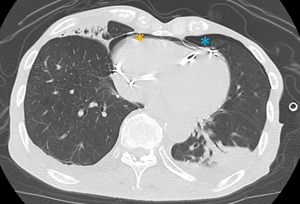
An 83-year-old man underwent dual-chamber pacemaker placement for complete atrioventricular block at another hospital. The active-fixation ventricular lead was positioned on the free wall of the anterior right ventricle. Ventricular pacing failure occurred on the day after pacemaker implantation, and fluoroscopy revealed right ventricular (RV) lead perforation. The patient was transferred to our hospital, and chest computed tomography revealed a severe pneumothorax and moderate pneumopericardium. These symptoms were relieved after chest tube drainage, and the patient’s hemodynamics stabilized. The RV lead was percutaneously removed using simple traction under fluoroscopic guidance with cardiac surgical backup and was uneventfully refixed to the RV septum. Although there have been several reports of pneumopericardium caused by atrial lead perforation, there are very few cases related to RV lead. Pneumopericardium complicated by pneumothorax due to RV lead perforation can be relieved using chest tube drainage without the need for pericardiocentesis.

Published
Issue
Section
License
Copyright (c) 2024 The authors

This work is licensed under a Creative Commons Attribution-NonCommercial 4.0 International License.


