A Rare Case of Cardiac Myxoma With Multiple Feeding Vessels From the Right Coronary Artery and the Left Circumflex Artery
DOI:
https://doi.org/10.14740/cr2113Keywords:
Myxoma, Neovascularity, Computed tomography coronary angiography, Left atrial thrombusAbstract
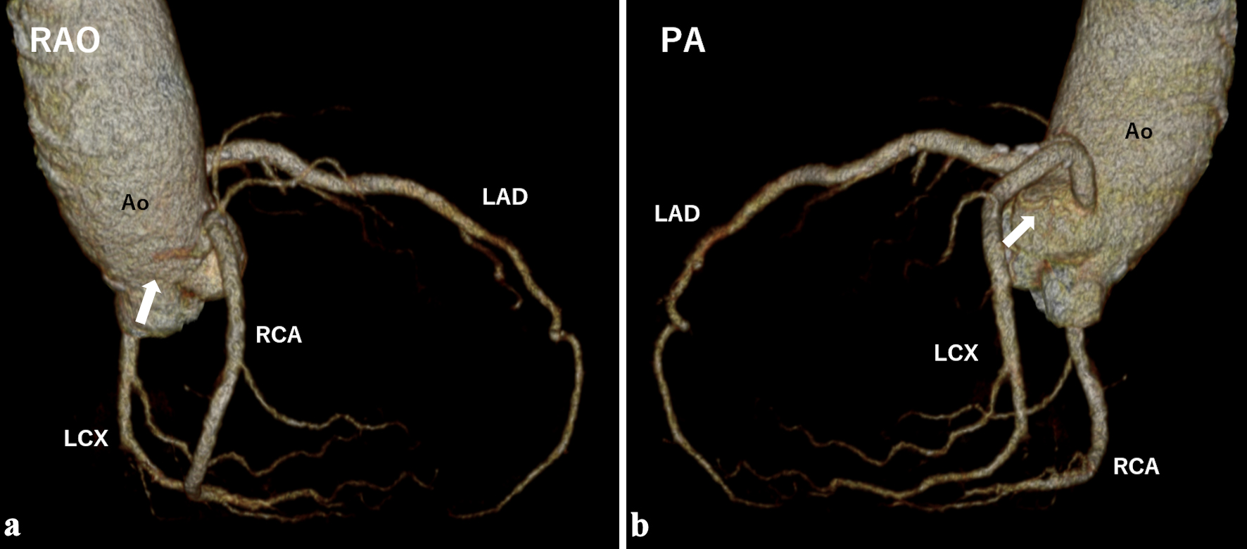
An 80-year-old woman with persistent atrial fibrillation was referred to our hospital for evaluation of a left atrial mass. Transthoracic and transesophageal echocardiography revealed a well-defined, sessile, and immobile mass attached to the interatrial septum. Computed tomography (CT) coronary angiography revealed a cardiac tumor fed by two vessels: one from the right coronary artery and one from the left circumflex artery. Based on these findings and cardiac magnetic resonance imaging, the mass was diagnosed as a left atrial myxoma, and excision was performed. Although some atrial myxomas are highly vascular, identification of multiple feeding vessels on CT coronary angiography is rare. Preoperative evaluation of feeding vessels is helpful in distinguishing myxomas from left atrial thrombi, especially in patients with hypercoagulability.
Published
Issue
Section
License
Copyright (c) 2025 The authors

This work is licensed under a Creative Commons Attribution-NonCommercial 4.0 International License.