


Left Ventricular Myocardial Deformation Abnormalities in Ankylosing Spondylitis (Radiographic Axial Spondyloarthritis): Association With Disease Severity Assessed by Two-Dimensional Speckle-Tracking Echocardiography
DOI:
https://doi.org/10.14740/cr2308Keywords:
Ankylosing spondylitis, Axial spondyloarthritis, Radiographic axial spondyloarthritis, Speckle-tracking echocardiography, Global longitudinal strain, Left ventricular dysfunction, Myocardial deformation, Cardiovascular involvement, Disease severity, SacroiliitisAbstract
Background: Ankylosing spondylitis (AS), also termed radiographic axial spondyloarthritis (r-axSpA) under current Assessment of SpondyloArthritis international Society (ASAS) nomenclature, is a chronic inflammatory disorder associated with an increased risk of cardiovascular involvement, including subclinical myocardial dysfunction. Conventional echocardiography often fails to detect early myocardial abnormalities despite preserved left ventricular systolic function. Two-dimensional speckle-tracking echocardiography (2D-STE) enables sensitive assessment of myocardial deformation through global longitudinal strain (GLS), facilitating the early detection of subclinical cardiac involvement. This study aimed to evaluate left ventricular myocardial deformation in patients with AS using 2D-STE and investigate its relationship with disease severity and duration.
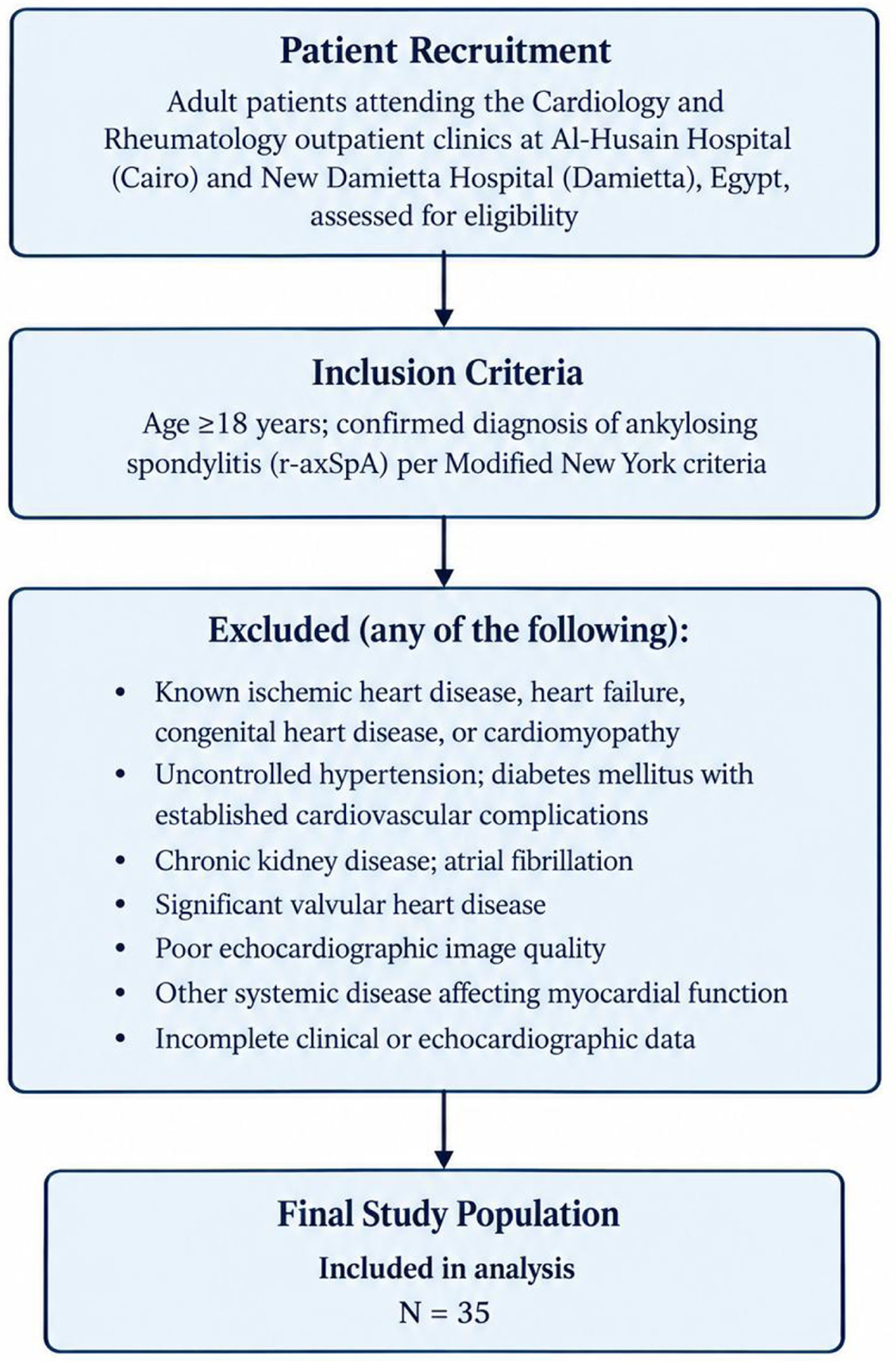
Methods: This cross-sectional observational study included 35 patients with AS diagnosed according to the Modified New York criteria. All participants underwent comprehensive clinical evaluation, laboratory investigations, conventional transthoracic echocardiography, and 2D-STE. Disease activity was assessed using the Bath Ankylosing Spondylitis Disease Activity Index (BASDAI) and Ankylosing Spondylitis Disease Activity Score (ASDAS). Radiographic disease severity was determined using the modified New York sacroiliitis grading system. Correlations between myocardial deformation parameters, radiographic severity, and disease duration were analyzed using Pearson’s correlation coefficient.
Results: The study population had a mean age of 33.9 ± 7.3 years, and 68.6% were male. Conventional echocardiography demonstrated preserved left ventricular systolic function, with a mean left ventricular ejection fraction of 65.6±5.0%. The mean GLS was −17.92±4.80%. Radiographic sacroiliitis grade demonstrated a significant positive correlation with global longitudinal strain (r = 0.345, P = 0.046), apical two-chamber longitudinal strain (r = 0.455, P = 0.008), and apical four-chamber longitudinal strain (r = 0.392, P = 0.022), whereas no significant association was observed with apical three-chamber strain, aortic root diameter, or valvular abnormalities. Disease duration showed a moderate negative correlation with GLS (r = −0.544, P = 0.001), indicating greater impairment of myocardial deformation in patients with longer-standing disease.
Conclusions: Patients with AS demonstrated evidence of subclinical left ventricular myocardial dysfunction despite preserved conventional systolic function. Greater radiographic disease severity and longer disease duration were significantly associated with impaired myocardial deformation assessed by speckle-tracking echocardiography. These findings support the potential role of GLS as a sensitive imaging biomarker for the early detection of cardiac involvement in AS and highlight the importance of cardiovascular surveillance in patients with advanced or longstanding disease.
Published
Issue
Section
License
Copyright (c) 2026 The authors

This work is licensed under a Creative Commons Attribution 4.0 International License.